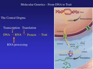
DNA
430 likes | 641 Vues
DNA. Discovery Structure Replication. 40 questions. 1. Describe the two strains of bacteria Griffith used in his experiment with mice. S strain (deadly) -produced a protective slime coating that helped it evade the mouse immune system. -caused pneumonia (a deadly lung disease) in

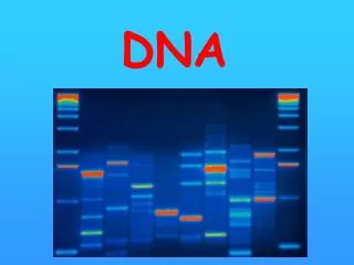
DNA
E N D
Presentation Transcript
DNA Discovery Structure Replication 40 questions
1. Describe the two strains of bacteria Griffith used in his experiment with mice. S strain (deadly) -produced a protective slime coating that helped it evade the mouse immune system. -caused pneumonia (a deadly lung disease) in mice. R strain (harmless) -did not produce a protective slime coating and therefore was easily defeated by the mouse immune system.
2. What happened to the mice when Griffith injected them with the heat-killed S strain? The mice lived.
3. What happened to the mice when Griffith injected them with a mixture of heat-killed S strain and live R strain? + The mice died of pneumonia.
4. A process in which one strain of bacteria is changed by a gene or genes from another strain of bacteria. Transcription Replication Transformation duplication
5. Oswald Avery used ____ to degrade (break down) various molecules taken form heat-killed bacteria. sulfur enzymes toxins x-rays
6. What types of macromolecules did Avery use enzymes on. Carbohydrates Proteins Lipids RNA DNA
7. What kind of enzyme did Avery use to degrade bacterial proteins? carbohydrase lipase protease DNAse Membrane protein
8. How did Avery ensure the validity of the results of his experiment with bacteria and mice? He degraded only one type of molecule at a time. He used all the enzymes at the same time. He decided not to degrade proteins and DNA. He injected a mixture of degraded molecules into mice at the same time.
9. Bacteriophages are a form of bacteria enzymes coils of DNA viruses
10. What two organisms did Hershey and Chase work with in their study of DNA? Bacteriophages and mice E. coli bacteria and mice Bacteriophages and E. coli bacteria Bacteriophages and viruses Bacteriophages E. coli bacterium
Bacteriophages Lysed Bacterium 11. Interpret this micrograph. Phages emerging Bacteriophages breaking out of a bacterium that has been infected.
12. What radioactive element did Hershey and Chase use to “tag” DNA? 32P (phosphorus) DNA
13. What radioactive element did Hershey and Chase use to “tag” the protein coat? 35S (sulfur) The amino acid methionine
14. Why can’t Hershey and Chase use to 35S to tag phage DNA? DNA does not contain sulfur.
15. What results did Hershey and Chase observe? The protein coats were injected into the bacterial cells causing transformation. Protein coats do not contain phosphorus. Radioactivity detected inside bacterial cells came from 32P and not 32S. Bacteriophages are good at infecting bacteria.
16. What can be concluded from the Avery and Hershey & Chase experiments? DNA is the transforming molecule. Proteins are larger than nucleic acids. Enzymes are good at breaking down molecules. The S-strain is more deadly than the R-strain.
17. Before DNA could be shown to be the genetic material in cells, scientists had to show that it could tolerate high temperatures carry, make copies of, and transmit information be modified in response to environmental conditions. be broken down into small subunits.
Phosphate group Nitrogenous base 18. A nucleotide does NOT contain a 5-carbon sugar. nitrogen base protein phosphate group Sugar (deoxyribose)
A C T G 19. According to Chargaff’s rule of base pairing, which of the following is true about DNA? Erwin Chargaff (1905 – 2002) = A = T, and C = G A = C, and T = G A = G, and T = C A = T = C = G =
20. Name the missing nitrogenous base. Thymine Guanine _____________ Cytosine Adenine
21. Use Chargaff’s rule to complete the table below. 30 ? + 60 100
21. Use Chargaff’s rule to complete the table below. 20 20 30 40 + 60 100
A C T G 22. The bonds that hold the two strands of DNA together come from The attraction of phosphate groups for each other. Strong bonds between nitrogenous bases and the sugar-phosphate backbone. Hydrogen bonds between nitrogenous bases. Carbon-to-carbon bonds in the sugar portion of the nucleotides.
A G C T T C A G 23. What is the term that describes how the two strands in DNA run in opposite directions? Antiparallel
24. Who took this photo? Photo 51 Rosalind Franklin (1920 – 1958)
25. What is this a photo ofand what technique was used to make it? The photo shows the structure of DNA Rosalind used X-ray diffraction to take the picture Photo 51
26. List two things that Rosalind Franklin learnedfrom her photo? DNA has a double helix shape DNA is made of two strands. The nitrogenous bases are near the center. Photo 51
27. Who are these two men and what are they famous for? James Watson and Francis Crick. They won the Nobel prize for building the first accurate model of DNA. James Watson (1928 - ) Francis Crick (1916 - 2004 )
A C B 28. Name the three parts of the nucleotide shown below? phosphate group sugar (deoxyribose) nitrogenous base
29. The process of copying DNA prior to cell division is called cytokinesis interphase base pairing replication
30. The diagram below shows the process of DNA replication digestion transformation transpiration
31. The enzyme that “unzips” DNA during replication is called DNA polymerase carbohydrase helicase replicase
32. The enzyme that “fastens” new nucleotides to the original DNA strand is called. carbohydrase DNA polymerase helicase replicase
33. In which direction is the circled DNA polymerase moving? Right to left
35. Is the chromosome shown below from a prokaryote or eukaryote? How do you know? It is from a prokaryote The DNA forms a loop or ring like this one
36. The micrograph below shows DNA in fruit flies. What are the “bubbles” (as indicated by the arrows) caused by? The bubbles are where DNA replication is taking place.
Replication nearly complete 37. Interpret the following sequence of diagrams? Replication in two directions Replication bubble The diagrams demonstrate prokaryote replication, which involves only one replication bubble. Replication proceeds within the bubble in opposite directions. Two identical chromosomes result
38. How many replication forks are shown in this micrograph? Replication forks There are two forks. One at each end of the bubble.
39. Is this a prokaryotic or eukaryotic chromosome? How can you tell? Human chromatids Eukaryotic The chromosome is rod shaped instead of circular.
40. What are the tips of chromosomes called and what enzyme replicates them. Telomeres Telomeres The tips are called telomeres The enzyme is telomerase