
Introduction
Thanks to Davidson College, NSF-REU, and Bank of America for their generous support. . Determining the Physical Properties of DNA in DNA Microarrays Using Optical Tweezers. Megan McDonald 1,2 , A. Malcolm Campbell 1 , Dan Boye 2. # 05-A-3139-BPS .

Introduction
E N D
Presentation Transcript
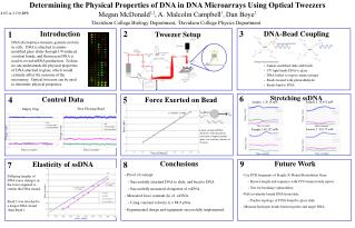
Thanks to Davidson College, NSF-REU, and Bank of America for their generous support. Determining the Physical Properties of DNA in DNA Microarrays Using Optical Tweezers Megan McDonald1,2, A. Malcolm Campbell1, Dan Boye2 # 05-A-3139-BPS 1Davidson College Biology Department, 2Davidson College Physics Department 3 2 1 Introduction DNA-Bead Coupling Tweezer Setup DNA microarrays measure genome activity in cells. DNA is attached to amino-modified glass slides through UV-induced covalent bonds, and fluorescent DNA is used to reveal mRNA production. To date, no one understands the physical properties of DNA attached to glass, which would certainly affect the outcome of the microarray. Optical tweezers can be used to determine physical properties. • Amino-modified slide and beads • UV light binds DNA to glass • DNA boiled to expose amino groups • Beads treated with glutaraldehyde • Beads bind to DNA Four quadrant 6 Stretching ssDNA 4 5 Control Data Force Exerted on Bead Sample 1: 20.18 mW Sample 2: 18.852 mW Empty Trap Free Floating Bead Voltage (volts) Voltage (volts) Force (pN) Time (seconds) Time (seconds) Sample 1 80.137 mW Sample 2: 80.137 mW Linear section of DNA elasticity, with maximum extension of approximately 4µm, at a constant velocity of 18 µm/s. Voltage (volts) Voltage (volts) Voltage (volts) Voltage (volts) Time (seconds) Time (seconds) Power (Watts) Time (seconds) Time (seconds) Conclusions 9 Future Work Elasticity of ssDNA 8 7 • Proof of concept: • Successfully attached DNA to slide, and bead to DNA • Successfully measured elongation of ssDNA. • Measured force constant (k) of ssDNA • Using constant velocity, k = 88.9 µN/m • Experimental design and equipment successfully implemented. • Use PCR fragments of Fragile X Mental Retardation Gene • Known length and sequence with CCG trinucleotide repeat. • Test for breaking vulnerability. • Pull covalently bound DNA from slide. • Predict topology of DNA bound to glass slide. • Measure hydrogen bonds between probe and target DNA. Differing lengths of DNA cause changes in the force required to stretch the DNA strand. Bead 2 was attached to a longer DNA strand than Bead 1. Time (seconds) Power (Watts) Helping Students Discover Genomics, Proteomics & Bioinformatics A. Malcolm Campbell, Laurie J. Heyer, Adam Abele, Brian Akin, Danielle Choi, Parul Karnik, Peter Lowry, David Moskowitz, Emily Oldham, Jennifer Madden


