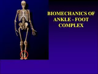
The Foot and Ankle Complex
The Foot and Ankle Complex. Sarah Rayner Extended Scope Practitioner Physiotherapist. Anatomy. The ankle and foot is a complex structure comprised of 28 bones (including 2 sesamoid bones) and 55 articulations (including 30 synovial joints), interconnected by ligaments and muscles

The Foot and Ankle Complex
E N D
Presentation Transcript
The Foot and Ankle Complex Sarah Rayner Extended Scope Practitioner Physiotherapist
Anatomy • The ankle and foot is a complex structure comprised of 28 bones (including 2 sesamoid bones) and 55 articulations (including 30 synovial joints), interconnected by ligaments and muscles • In addition to sustaining substantial forces, the foot and ankle serve to convert the rotational movements that occur with weight bearing activities into sagittal, frontal, and transverse movements
Anatomy : Foot • Hindfoot (posterior segment): talus and calcaneus • Midfoot (middle segment): navicular, cuboid and 3 cuneiforms • Forefoot (anterior segment): metatarsals and the phalanges
Anatomy: Surface marking practical • Talocrural joint line • Medial malleolus • Lateral malleolus • Navicular • 1st MTP joint • Achilles tendon • Tibialis posterior tendon • Anterior talofibular ligament • Calcaneofibular ligament • Peroneus longus and brevis • Plantarfascia attachment to calcaneus • Midtarsal joint line
Conditions: lateral ligament injury • Acute inversion of ankle • Usually occurs in sports requiring quick change of direction especially if it takes place on uneven surfaces such as grass. • Also common in sports when a player has jumped and lands on top of another players feet. • Most common mechanism is Inversion coupled with PF. • ATFL injured first then CFL as ATFL is taut in PF On Examination: • Lateral ankle pain and swelling • Pain on inversion combined with plantarflexion • Tests: Anterior draw and talar tilt
Conditions: lateral ligament injury • Management • PRICE • Graded return to sport • May require Physiotherapy • Rate of recovery dependent on severity • Failure to resolve • Continued instability or possible OCD • Refer to CATTS / Orthopaedics • May require further investigations ? MRI • Surgical intervention (arthroscopy +/- stabilisation procedure
Conditions: Plantarfasciitis • Insertional heel pain of the plantar fascia with or without a heel spur. • Biomechanical abnormalities cause pathological stress to the plantar soft tissues • Typical presentation: • Isolated heel pain on initiation of WB (on rising am or after prolonged sitting/rest) • Predisposing factors: • High BMI • Tightness of TA • Inappropriate shoe wear • On Examination • Pain on palpation at plantar fascia insertion
Conditions: Plantarfasciitis management • Initial self directed treatment (up to 6 weeks): • NSAID’s • Regular calf and plantar fascia stretches • Avoidance of flat shoes and barefoot walking • OTC arch supports and heel cushions • Ice • Weight loss • Limitation of extended physical activity • Consider steroid injection where appropriate • If failing to improve refer on to local CATTS/MSK service: • Custom orthotics (podiatry) • Night splints • Steroid injections • Immobilisation • Extracorpeal shockwave therapy • Surgical plantar fascia release
Conditions: Achilles tendinopathy • Non-insertional: • Usually a degenerative mid substance lesion • Often with neovascularisation and proliferation of neural structures in the area which cause pain • Often poor collagen structure, poor healing and no inflammation on imaging • Insertional: • Change in microscpic structure with increased Glycosaminoglycans • Change in fibrillar structure giving swelling • Tendinitis / tendinosis depends on degree of inflammation • Bursitis often associated with Haglund’s deformity (“pump bumps”)
Conditions: Non-insertional Achilles Tendinopathy • Presentation: • Most common in males but seen in all ages • Pain on Achilles loading (walking, running) • Can be debilitating • Fusiform swelling • Tightness of Gastrocnemius • Treatment: • Eccentric loading exercises • Stretches • Correct abnormal biomechanics • Physiotherapy / podiatry • Extracorpeal shockwave therapy
Conditions: Insertional Achilles Tendinopathy • Management • Initial conservative treatment as for non-insertional Achilles tendinopathy • Surgical debridement
Conditions: Achilles Ruptures • Presentation: • Patients usually feel POP in Achilles area • POP may be heard • Usually occurs in the avascular area of the Achilles 5 – 10cm above the insertion • Common in Badminton , Squash and football in that order • Usually occurs to the end of a game • On Examination: • +ve calf squeeze • Palpable dip • Management • Surgical
Conditions: Ankle Impingement • Anterior bony impingement: • Pain usually over anterior ankle • Pain may be anterolateral • Osteophytes usually palpable and may be associated with loss of ROM particularly dorsiflexion • Arthroscopy • Posterior Impingement • Os trigonum, Bony osteophytes • Adhesions, synovitis ; Multiple injuries or hypermobility (dancers) • FHL tendinitis • Subtalar impingement • If conservative treatment fails, posterior ankle arthroscopy
Conditions: Tibialis Posterior Dysfunction • Common cause of acquired flatfoot in adults • Women over 40 most at risk • Presenting features: • Pain and swelling medial hindfoot • Change in foot shape reported • On Examination: • Valgus heel, flattened longitudinal arch and abducted forefoot • Pain on resisted inversion and on palpation tibialis posterior • Pain and dysfunction on single leg heel raise
Conditions: Tibialis Posterior Dysfunction Management • Conservative treatment • Rest • Orthotics and podiatry • Weight management • Surgical management • Hindfoot osteotomy with tendon transfer • Arthrodesis of the hindfoot
Conditions: Hallux Rigidus • 1st MTP Arthritis • Epidemiology: • Women > men • 60% bilateral • Late adulthood • Etiology: • Direct: trauma, fracture • Indirect: TMT hypermobility, flat 1st MTP joint, Long 1st MT, pesplanus, inflammatory • Clinical Symptoms: • Limited 1st MTP movement • Pain on toe off • Pain with activity • Pain with shoewear • Swelling • Limp: lateral foot WB, external rotation of hip
Conditions: Hallux Rigidus • Management: • Conservative • Footwear • Activity modification • Podiatry • Injections • Surgery • Cheilectomy • Osteotomy • Joint replacement • Fusion
Conditions: Morton’s Neuroma • Swelling of nerve and scar tissue arising from compression of the interdigital nerve • Often pain radiating into the toes accompanied by pins and needles • Pain increased by forefoot weight bearing and with narrow fitting footwear • On Examination: • Interdigital pain commonly in the 3rd and 2nd interdigital space • +ve Mulder’s test • Management: • Orthotics • Injection • Surgical removal
Examination: Summary • As always take a good history to guide your examination: site of pain, overuse or trauma, swelling, WB status etc. • Gait and function (heel raise, weight transfer, proprioception) • Observations: in standing and sitting/lying • Swelling, heat, scars, bruising, circulation, deformity • Biomechanics (pronation/supination, abducted) • ROM • Resisted testing • Palpation • Special Tests • Anterior draw rest • Talar tilt test • Squeeze test • Calf squeeze test (Thompson test) • Lateral squeeze test for Morton’s neuroma (Mulder’s click)
Case Studies: Practical • Monica a 30-year-old medical receptionist presents with sore Achilles tendons. Over the weekend she has done a 15-mile sponsored walk. She is a bit annoyed because although she does not do any significant walking she feels that she keeps herself very fit with her Latin American dancing. She also bought an expensive pair of Nike trainers especially for the walk. • A 45-year-old lady complains of pain in her right heel. This started 3 weeks ago after she had spent the weekend helping her husband lay some flags for a patio. She describes how it feels as if she has a small ball bearing under her heel when walking. • A 65-year-old man complains of gradually increasing pain in the ball of his right foot over several months. He has had to curtail his ballroom dancing and of late his walking is becoming restricted. • A 13-year-old girl who enjoys ballet is finding increasing pain in her left big toe with her dancing. She says her big toes are not straight anymore. • A 46-year-old farmer complains about his left ankle. Apparently a year ago he had a "bad sprain" when he inverted the ankle as he was trying to catch a sheep. He went to casualty and had an X-ray (NBI) and came away with a tubigrip bandage. He was not followed up. Since then he finds himself "going over" on the ankle on uneven ground if he is not watching carefully where he puts his feet. The ankle is frequently swollen following these episodes.
Any Questions? Thank you