Practical Of Genetics
Practical Of Genetics. Lab 4 & 5 Human Karyotype. Objectives:. 1. Students will be able to demonstrate a microtechnique for reliable chromosomal analysis of leucocytes obtained from peripheral blood.

Practical Of Genetics
E N D
Presentation Transcript
Practical Of Genetics Lab 4 & 5 Human Karyotype
Objectives: 1. Students will be able to demonstrate a microtechnique for reliable chromosomal analysis of leucocytes obtained from peripheral blood. 2. Students will be able to prepare a karyotype from the chromosomes of a normal human male or female. 3. Students will be able to use the karyotyping techniques for diagnosing a chromosomal disorder.
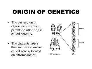
INTRODUCTION • Chromosomal aberrations are abnormalities in the number or microscopically observable structure of chromosomes. • The “normal” human diploid cell has 23 pairs of chromosomes visible only at the metaphase stage of mitosis, 22 homologous pairs of autosomesand two sex chromosomes.
Each chromosome has a characteristic size and shape in the “normal” cell. • Chromosome pair #1contains the largest chromosomes, while the Y chromosome is the smallest. • Chromosomes of similar size and morphology are grouped together by letter; the chromosomes can be arranged in 7 groups (A, B, C, D, E, F, G). • Thus group A contains pairs #1, #2, and #3 chromosomes.
A chromosome is divided by its centromer into short arm p (= petite or short) and long arm q (= queue, or long) p q
Chromosomes can be classified by the position of their centromer:
Telocentric Centromere is located at the terminal end of the chromosome. • Humans do not possess any telocentric chromosomes.
Chromosomes are arranged into seven groups A1-3 Large metacentric B4,5 Large submetacentric C6-12, X Medium submetacentric D13-15 medium acrocentric E16-18 short metacentric F19-20 Short metacentric
Karyotypingis a technique that allows for the visualization and identification of human metaphase chromosomes.
Samples for chromosomal analysis can be prepared relatively easily using skin, bone morrow, chorionic villy or cells from amniotic fluid.
It is estimated that one in 156 live births have some kind of chromosomal abnormality. • To create a karyotype, chromosomes from a cell are arranged, stained and photographed. • The photograph is enlarged and cut up into individual chromosomes. • The homologous chromosomes can be distinguished by length and by the position of the centromer.
A blood sample is taken and white blood cells grown in special medium for three days under the influence of the mitotic stimulant ( phytohemagglutinin " PHA " ) to enter into mitosis by DNA replication.
After 68-72 hours, amitotic inhibitor ( Cholchicin) is added to the culture to stop mitosis in the metaphase stage. • After treatment by hypotonic solution ( KCl ) to causes a swelling of the cells and allow dispersion of the chromosomes within the cell membrane.
Fixation is used as wash solution to lyse the remeaning red cells and remove some chromosomes protein. • After staining, chromosomes can be microscopically observed and evaluated for abnormalities.
Metirals • Heparinzed whole blood • Heparin sodium injection • Peripheral blood Karyotyping medium with PHA (RPMI 1640) • Incubator 5% CO2 at 37 °C • Colcemide solution 10g/ml • 0.075 M KCl • Fixative solution ( 3x methanol : 1x glacial acetic acid ) • Giemsa stain solution • Slides and Microscope
Peripheral blood media preparation • Blood culture media; • 500 ml RPMI 1640with 100mlfetal bovine serum, 6.5ml penicillin– streptomycin and 7mlglutamine. • Dispense 10ml aliquots into sterile tube and add 2% (0.2ml) PHA to each tube. • Store at 4C for along as 2 weeks.
Stains and Dyes • Used to produce a pattern of bands specific to each type of chromosome • One common method is G-banding • Treated with trypsin • Stained with Giemsa stain
Karyotyping procedure 1- Inoculate 0.5ml of heparinized whole blood into tube with 10ml of karyotyping medium. 2- Incubate the tubes in incubator with 5% CO2 at 37 oC for total of 72 hours. 3- After total of 69 hours from seeding add 100μl of Colcemid Solution to each culture tubes. 4- Incubate the tubes at 37 oC for an additional 20-30 minutes. 5- Spin at 500g (1500 rpm) for 7 minutes. 6-Remove the supernatant and re-suspend the cells in 5ml of hypotonic 0.075M KCl prewormed to 37oC.
7- Incubate at 37oC for 15 minutes. 8- Add drop-by- drop (with vortexing) 1ml fresh ice cold fixative. 9- Spin at 500g (1500RPM) for 7 minutes. 10- Remove the supernatant, agitate the cellular sediment and add drop-by- drop (with continous vortexing), 5ml of fresh, ice-cold fixative. 11- Leave at 4ºC for 20 minutes. 12- Repeat steps 9 and 10,until the supernatant is clear. 13- Spine at 500g (1500RPM) for 7 minutes. 14- Re-suspend the cell pellet with a 1.5ml of fresh fixative.
15- Drop 4-5 drops, from ahigh of approximately 30cm onto aclean slide and blow carefully on the drops for spreading them on the slide. 16- Put the slides on a 45ºC heated plate for 2-4 minutes. 17- Heat the slides to 60 ºC for overnight or to 90 ºC for 90 minutes. 18- Place the slides and flood them with Giemsa stain solution for 8 minutes. 19- Gently rinse the slides in distilled water and air dry. 20- Observe the chromsomes under microscope by using 10, 40 and 100x and photograph it and cut each chromosome from the photograph and arrange the chromosomes according to the size and position of centomer.
Staining procedure Materials: 1.Giemsa stain solution - 6ml Giemsa stain in 70ml ddH20 2.HBSS 3.Trypsin X10 4.PBS without Ca, Mg
Method 1. Incubate the slides in Trypsin solution:2ml Trypsin x10 in 50ml of HBSS at room temperature. 2.After 2.5 min., neutralize the trypsin by immersing the slides in PBS with 5% FCS. 3. Rinse the slides in PBS. 4.Place the slides horizontally and flood them with stain solution for 2.5 minutes. 5.Gently rinse the slides in ddH20 and air-dry. • Exact times of Trypsin treatment and staining should be determined personally by each lab to get optimal results
Chromosomes are arranged by : • Size. • Position of the centromere. • Specific banding patterns.