Syphilis
Syphilis. Boris Ioffe, D.O. Syphilis. AKA lues Contagious, sexually transmitted disease caused by the Spirochete: Treponema pallidum Enters through skin or mucous membrane where primary manifestations are seen. Treponema pallidum. Spiral spirochete that is mobile


Syphilis
E N D
Presentation Transcript
Syphilis Boris Ioffe, D.O.
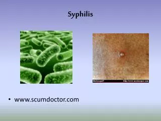
Syphilis • AKA lues • Contagious, sexually transmitted disease caused by the • Spirochete: Treponema pallidum • Enters through skin or mucous membrane where primary manifestations are seen
Treponema pallidum • Spiral spirochete that is mobile • # of spirals varies from 4 to 14 • Length is 5 to 20 microns • Can be seen on fresh primary or secondary lesions by darkfield microscopy or fluorescent antibody techniques
Syphilis epidemiology • Major health problem throughout world • 2.6 cases per 100,000 in 1999 in the US • Lowest level ever recorded • Concentrated in 28 counties in the SE U.S. • Mainly gay men and crack cocaine users
Syphilis epidemiology • Enhances risk of transmission of HIV • HIV testing recommended in all patients with syphilis • Reportable disease
Serologic Tests • Testing reveals patients immune status not whether they are currently infected • Non-treponemal antigen test uses lipoidal antigens rather than T. pallidum or components of it • RPR = rapid plasma reagin • VDRL = Venereal Disease Research Laboratory
Serologic Tests • Positive within 5 to 6 weeks after infection • Strongly positive in secondary phase • Strength of reaction is stated in dilutions • May become negative with treatment or over decades
Serologic Tests • MHA-TP: microhemagglutination assay for T. pallidum • FTA-ABS: fluorescent treponemal antibody absorption test • All positive nontreponemal test results should be confirmed with a specific treponemal test
Serologic Tests • Treponemal tests become positive early, useful in confirming primary syphilis • Remain positive for life, useful in diagnosing late disease • Treatment results in loss of positivity in 13-24% of patients
Biologic False-Positive Test Results • Positive test with no history or clinical evidence of syphilis • Acute BFP: those that revert to negative in less than 6 months • Chronic BFP: those that persist > 6 months
Acute BFP Vaccinations Infections pregnancy Chronic BFP Connective tissue disease (SLE) Liver disease Blood transfusions IVDA BFP Test Results in Syphilis
Cutaneous Syphilis • Chancre is usually the first cutaneous lesion • 18 to 21 days after infection • Round indurated papule with an eroded surface that exudes a serous fluid • Usually painless and heals without scarring
Chancre • Inguinal adenopathy 1-2 weeks after chancre • Generally occur singly, but may be multiple • Diameter mm to cm
Chancres • In women, the genital chancre is less often observed due to location within the vagina and cervix • Edema of labia may occur
Chancre • Untreated, the chancre heals spontaneously in 1 to 4 months • Constitutional symptoms begin just as chancres disappear • Extragenital chancre: may be larger, frequently on lips, rarely tongue, tonsil, breast, finger, anus.
Chancre Histology • Ulcer covered by neutrophils and fibrin • Dense infiltrate of lymphocytes and and plasma cells • Spirochetes seen with with silver stains; Warthin-Starry • Direct fluorescent antibody tissue test (DFAT-TP) = serous exudate collected on a slide sent for exam
Serology • Nontreponemal tests positive 50% • Treponemal tests positive 90% • Positivity depends upon duration of infection, if chancre has been present for several weeks, test is usually positive
Incubation 3 weeks Painless Hard Lymphadenopathy may be bilateral, nontender, nonsuppurative Incubation 4-7 days Painful Soft Lymphadenopathy unilateral, tender, suppurative Chancre vs. Chancroid
DDx in Syphilis • Chancroid - multiple lesions, may coexist with chancre, must r/o syphilis • Granuloma Inguinale - indurated nodule that erodes, soft red granulation tissue, Donovan bodies in macrophages with Wright or Giemsa stain • Lymphogranuloma Venereum - small, painless, superficial non indurated ulcer, primary lesions followed in 7 to 30 days by adenopathy • HSV - grouped vesicles, burning pain
Secondary Syphilis • Skin manifestations in 80% called syphilids • Symmetric, generalized, superficial, macular - later papular, pustular • May affect face, shoulders, flanks, palms and soles, anal or genital areas
Secondary Syphilis Macular Eruptions • Exanthematic erythema 6-8 weeks after chancre - may last hours to months • Round, slightly scaly ham-colored macules • Pain and pruritus may be present • Generalized adenopathy
Secondary Syphilis Papular Eruptions • Occurs on face and flexures of arms, legs, and trunk • Yellowish-red spots may appear on palmar and plantar surfaces • Ollendorf’s sign = tender papule
Secondary Syphilis Papular Eruptions • May produce a psoriasiform eruption • May appear as minute scale-capped papules • Tend to be disseminated, but may be localized, asymmetrical, configurate, hypertrophic or confluent.
Secondary SyphilisPapular Eruptions • Annular syphilid - mimics sarcoidosis and is more common in blacks • Pustular syphilid – rare - face, trunk, extremities red small crust-covered ulceration • Rupial syphilid - superficial ulceration is covered with a pile of terraced crusts resembling an oyster shell.
Secondary SyphilisPapular Eruptions • Lues Maligna - rare, severe ulcerations, pustules, or rupioid lesions, accompanied by severe constitutional symptoms. • Condylomata lata - papular mass, weeping, gray 1-3cm, groin, anus (not vegetative like condylomata acuminata) • Syphilitic alopecia - irregular, scalp has a moth-eaten appearance 5% of pts
Secondary SyphilisMucous Membrane • Present in 1/3 of secondary syphilis • Most common is “syphilitic sore throat” • Diffuse pharyngitis, hoarseness • Tongue may show patches of desquamation of papillae • Ulcerations of tongue and lips in late stages
Secondary SyphilisMucous membrane • Mucous patches are the most characteristic mucous membrane lesions; macerated, flat. Grayish, rounded erosions covered by a delicate, soggy membrane.
Secondary SyphilisSystemic Involvement • Lymphadenopathy common. • Acute glomerulonephritis, gastritis, proctitis, hepatitis, meningitis, iritis, uveitis, optic neuritis, Bell’s palsy, pulmonary nodular infiltrates, osteomyelitis, polyarthritis.
Secondary SyphilisDiagnosis • Nontreponemal serologic tests for syphilis are strongly reactive (seronegativity rarely in AIDS) • Spirochetes on darkfield exam
Pityriasis rosea Drug eruptions (pruritic) Lichen planus; Wickham’s striae, Koebner’s, pruritic Psoriasis; no adenopathy Sarcoidosis; need serology and silver staining of biopsy Infectious mononucleosis, false pos RPR Geographic tongue Aphthous stomatitis Secondary SyphilisDDx “Great Imitator”
Latent Syphilis • After the lesions of secondary syphilis have involuted, a latent period occurs where the patient has no clinical signs, but positive serological tests • May last a few months or a lifetime • 60-70% of pts that are untreated remain asymptomatic for life • Women may infect unborn child for 2 years
Late Syphilis • Defined by CDC as infection of greater than 1 years duration • Tertiary Cutaneous Syphilis • Late Osseous Syphilis • Neurosyphilis • Late Cardiovascular Syphilis
Tertiary Cutaneous Syphilis • Tertiary syphilids usually occur 3-5 years after infection • 16% of untreated pts will develop lesions of skin, mucous membrane, bone or joints • Skin lesions are localized, destructive, heal with scarring
Tertiary Syphilids • Two main types; Nodular syphilid and the Gumma • Nodular - reddish brown firm papules or nodules 2mm or larger, scales. • Gumma - larger
Nodular Tertiary Syphilid • Lesions tend to form rings and undergo involution as new lesions develop • Characteristic circular or serpiginous pattern • “kidney-shaped” lesion occurs on the extensor surfaces of the arms and on back • Patches have scars and fresh ulcerated lesions • Process may last for years, slowly marching across large areas of skin
Gumma • May occur as unilateral, isolated, single or disseminated lesions, or serpiginous • May be restricted to the skin, or originate in deeper tissues, and break down the skin • Lesions begin as small nodules, enlarge to several centimeters • Central necrosis, deep ulcer with a gummy base, most frequent site is lower legs