Download
1 / 43
620 likes | 1.37k Vues
Tibial Plateau Fractures. Roman Schwartsman M.D. Las Vegas Nevada. Classification - AO. Classification - Schatzker. Why Ilizarov?. Biomechanical advantage 4 wires & a screw are as strong as two plates (T. Watson) Fibular head wire will increase stability up to 30 %. Indications. Open Fx

E N D
Tibial Plateau Fractures Roman Schwartsman M.D. Las Vegas Nevada
Biomechanical advantage4 wires & a screw are as strong as two plates(T. Watson)Fibular head wire will increase stability up to 30%
Indications • Open Fx • Degloving injury • Compromised skin • Severe comminution • Shaft extension
Objectives • Joint congruity • Axial alignment • Stable fixation • Functional ROM & WB • Soft tissue healing
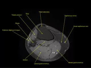
Pre-op evaluation • Skin & soft tissues • Radiographs • CT scan
Fracture topography • Condylar comminution • Tibial tubercle Fx. • Posterior cortex • Metaphysis, shaft
Fracture reduction • Skeletal traction • Closed reduction • Percutaneous reduction • Open reduction
Reduction first(or possibility of reduction)- Articular surface- Metaphysis, ShaftFrame applicationafter
Tibial plateauMedial Lateral Larger Smaller Stronger Weaker Higher Lower Concave Convex Meniscus small Meniscus large
Wire placement • 3-point fixation (in 3 planes: coronal, axial, saggittal) • Olive wires only • Push olive wires • Pull olive wires
3 1 4 2 Drop wire 2 MedialFx.
1 4 Drop wire 2 3 Anterolateral Fx.
5 3 4 1 2 Coronal split - reduce the split first
Pull wire Drop wire Bicondylar comminution with pull wire to avoid clustering on the ring
Safe wire placement(JOT May 1999 Thomas A.DeCoster et al )-safe corridors for wire placement-extent of capsular attachment
Capsular attachment Zone I - less than 6mmZone II - up to 12 mmZone III - up to 30 mmZone IV-12mm (8-14 mm)
-Other points: -10% of cases, the knee joint communicates with the proximal tibiofibular joint -pes anserinus bursa: a symptomatic wire
Femoral ring • Severe comminution (AO - B3,C3) • Articular surface inst. • Metaphyseal – diaphyseal separation (Schatzker –VI) • Ligamentous instability
Complications • Fx. related • Surgeon related • Patient related
Complications • Subchondral collapse • Technical errors Poor Fx reduction Wrong wires Wrong frame Instability • Patient compliance
Conclusions It is up to you… In our hands the best results are with closed reduction and Ilizarov frame application…
Thank you! LasVegasOrtho.com