Nanobiotechnology Introduction
Nanobiotechnology Introduction. Fall 2011 Graduate School of Biotechnology. Nanometer. :. =. :. What is Nanotechnology?.

Nanobiotechnology Introduction
E N D
Presentation Transcript
NanobiotechnologyIntroduction Fall 2011 Graduate School of Biotechnology
Nanometer : = :
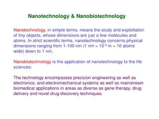
What is Nanotechnology? • “Nanotechnology is the understandingand control of matter at dimensions of roughly 1 to 100 nanometers, where unique phenomena enable novel applications. Encompassing nanoscale science, engineering and technology, nanotechnology involves imaging, measuring, modeling, and manipulating matter at this length scale.” Courtesy from National Nanotechnology Initiative http://www.nano.gov/html/facts/whatIsNano.html
Is Nanotechnology New? • Not really, other names used in the past: • Chemistry • Biochemistry • Microbiology • Atomic and Molecular Physics • Molecular Chemistry, Molecular engineering • It has been around for centuries
Nanoscience vs Nanotechnology • Nanoscience is concerned with the study of novel phenomena and properties of materials that occur at extremely small length scales. • Nanotechnology is the application of nanoscale science, engineering and technology to produce novel materials and devices. MANCEF Roadmap 2nd Edition
Fullerenes A geodesic sphere The Montreal Biosphere by Buckminster Fuller 1967 • Fullerenes are a recently discovered form of carbon by Kroto, Curl, and Smalley and then named after Buckminster Fuller. They can take the form of a hollow sphere, ellipsoid, or tube. Spherical fullerenes are sometimes called buckyballs, the C60 variant is often compared to a typical black and white football. Cylindrical fullerenes are called buckytubes. Fullerenes are similar in structure to graphite, which is composed of a sheet of linked hexagonal rings, but they contain pentagonal (or sometimes heptagonal) rings that prevent the sheet from being planar. • Application include cosmetics and pharmaceuticals, lubricant for engine parts, and electronics. • Eg.> anti-aging facial creams, Zelens in London
Carbon Nanotube (CNT) • Discovered 1991, Iijima • Armchair SWCNT • Zigzag SWCNT • Chiral SWCNT • STM image of 1.3nm-diameter chiral SWCNT
Same element with different property CdSe single crystal CdSe quantum dot
Same element with different property Emission spectra of Q-dot of various sizes
Preparation of Nanostructures Macroscopic or Mesoscopic Materials “Top down” approach “Top down” approach Microstructures (>100 nm) “Bottom up” approach Nanostructures (1-100 nm) “Bottom up” approach Molecules
“Top down” Miniaturization Photolithography • Photolithography from latin: light stonewriting • Photolithography is the process of transferring geometric shapes on a mask to the surface of a silicon wafer • The steps involved in the photolithographic process are: • wafer cleaning • barrier layer formation • photoresist application • soft baking • mask alignment • exposure and development • hard-baking.
From ENIAC(1944) to Tukwila (2008) • Weigth: ca. 30 t N. • Elements: 19000 tubes • Consumption: 200 kW 21 x 32 mm2 N. elements: 2 billions of transistors (Itanium -2GHz) Power consumption: ca. 170 W Elements size (min): ca. 65 nm
“Bottom up” Strategy – Molecule by Molecule Molecular recognition Selective binding of a substrate by a receptor Energy (Interaction) Information (Selection) • For effective molecular recognition, receptor and substrate should exhibit • Steric complementarity (shape and size) • Interactional complementarity • Large contact area • Multiple interaction sites • Strong overall binding
Cellular Components: A Self-Assembled Nanostructure
In the intersections of nanotechnology and biology • Nanotechnology offers biology new tools • Biology offers nanotechnology access to new types of functional nanosystems
The story is not entirely ‘nano’ but includes structures having a wide range of size • When small structures are considered for biological applications, or when small biologically derived structures are determined to have remarkable properties, the size of the system can be ‘nano (which we define, with some arbitrariness, as 1-100 nm) but also ‘micro’ (from 100 nm, or 0.1 um, to perhaps 1,000 um, or 1 mm)
The range of size covered by these terms-nanoscale, microscale and simply ‘small’-is important: structures vital to the cell have dimensions ranging from those of small molecules to those of millimeter-scale fluidic devices; which size is most important depends on what question one is asking. Enthusiasm for the potential value of ‘nano’ should be balanced against the established and rapidly expanding value of ‘micro.’
Much of the initial impetus for the development of nanotechnology came from its relevance to electronics. Microelectronics has, unarguably, changed the world through its impact on information processing and communications, and the progress in the field has been measured by its adherence to Moore’s Law: as microelectronic devices have become smaller, they have become less expensive, faster and more portable.
The situation in biological science is different for several reasons. First, biological structures are relatively large compared to structures in electronics and in physical nanoscience. The technological imperative to make things smaller that has dominated microelectronics for the past 50 years does not have the same urgency in biology.
The situation in biological science is different for several reasons. Second, the scanning probe microscope has been less revolutionary in its impact on biology than on the physical sciences. Biological structures-even those on surfaces-are soft and electrically insulating, and not easily imaged. Moreover, most of biology goes on inside the cell where the scanning probe tip cannot reach
Molecular machines and organelles The ultimate in functional nanosystems—‘biological nanomachines’—populate the cell. The ribosome, Na+/K+ ATPase, flagellar micromotor of bacteria, linear micromotors of muscle and of the microtubules that organize and move the cell, voltage-gated ion channels, DNA replication complexes, multimeric membrane receptors, and the photosynthetic reaction center: these, and countless other structures in the cell, are astonishingly complex, nonintuitive in their operation, and instructive to contemplate.
Molecular machines and organelles (cont.) Understanding biological nanostructures will be enormously stimulating for nanotechnology. The concept of very tiny machines has always appealed—to scientists and nonscientists alike—but the nascent field of nanotechnology originally assumed that nanomachines would probably resemble macromachines in their design. It is seldom possible to prove that something cannot happen in science, and it is difficult to prove that one cannot build nanomachines that are analogous to familiar macromachines. The fact that biological structures—although functioning in familiar ways—operate using principles that are entirely unfamiliar based on everyday experience suggests that would-be designers of nanomachines have much to learn from biology.
Molecular machines and organelles (cont.) An example is the rotary flagellar motor of bacteria (and the functionally related Na+/K+ ATPase). This motor does have a structure that serves as a shaft, and another structure that anchors the motor in the cell membrane, but beyond that, any resemblance to a macroscopic motor (whether internal combustion or electrical) stops. The components of the machine are complex, three-dimensional structures (proteins) that self-assemble in a series of steps, starting from a linear polypeptide chain; the mechanism of rotary motion seems to involve a sequential set of changes in conformation of the proteins driven by ions moving across the cell membrane. It certainly does not involve electrical current, magnetic fields or expansion of hot gasses in a cylinder.
Nanotips and microspheres The origin of nanotechnology is tools for imaging nanostructures: originally electron microscopy, but more recently, and famously, scanning probe devices. Although the direct imaging of biological structures has proved difficult, scanning probe devices have been enormously successful in allowing the application of forces directly to single molecules. The kind of information provided by these force-distance curves has, for example, made it possible to infer how stress is stored as strain in molecules by the unfolding of protein domains. Complementary information comes from studies in which the force is applied using optical tweezers or more recently, magnetic beads. The use of optical tweezers has provided insights into the mechanism and function of proteins involved in active transport in the cells (e.g.,myosin and kinesin)
Optical tweezer Steven Chu
Channels and pores Microchannels are the basis of microfluidic devices. When microchannels were made in silicon using conventional photolithography, the techniques were too slow, specialized and cumbersome to be broadly useful to biologists. The development of softlithographic methods for microfabrication, and the realization that high-resolution printing provided adequate resolution for fabrication of channels with widths from 10 μm to 1 mm, opened the door to active development of this area as a convenient and practical technique for fabricating microfluidic devices. The physics of fluids flowing in microchannels provided another important component of this area of microtechnology. In microchannels, aqueous buffers almost always flow laminarly—that is, without eddies, and with only diffusive mixing of streams flowing side by side. This combination of physical phenomena has begun to generate a wide variety of new devices: micro cell separators and particle counters, microsystems for cell culture, gradient generators and analytical systems.
Generation of gradients in a microfluidic device. The photograph shows a microfluidic device that generates a gradient of green and red dyes in solution. The three incoming channels (left) were connected to syringes via tubings (not visible). After the streams were combined into a single, wide channel (right), a gradient was formed across the channel, perpendicular to the direction of flow.
Channels and pores (cont.) Micro- and nanofabrication techniques also offer access to pores (which are, in essence, very short channels). Nanofabricated pores with dimensions down to approximately 2 nm have been demonstrated and proposed as the basis for single-molecule DNA sequencing. Interestingly, biology is beginning to offer its own set of methods for making channels and pores with nanoscale dimensions; the ease with which some of these systems can be assembled suggests that they may, in the long run, provide systems that are at least as useful as those fabricated top-down. Lipid nanotubes and channels in lipid membranes based on pore-forming peptides and proteins are examples.
Nanoparticles Nanoparticles are among the first nanoscale materials to be directly useful in biology: fluorescent particles labeled with antibodies (as tags that do not photobleach) and superparamagnetic magnetite particles coated with dextran (as imageenhancement agents in magnetic resonance imaging) are commercially available; a wide range of other fluorescent or magnetic nanoparticles will soon be available. Eventually, these small particles must be made much more useful and informative if they are to play an important role in understanding the workings of the cell.
Conclusions • Physical and biological sciences show nanoscience in different lights. • The road from reading the information in the genome to understanding life will be an arduous one, and reading the genome may be the easiest part • Understanding how molecules organize and function in cells will require new tools and concepts, and as the assemblies of molecules of greatest interest will have nanometer dimension, the tools must be appropriate for the task: that is, they must be able to characterize structures 0.1~0.001 times the size of the cell
Conclusions (cont.) • Selecting, sorting, maintaining, stimulating, herding and characterizing the cells will require tools substantially larger than the cell. Both nanometer and micrometer dimensions are relevant. • Nano-and microscience will show different aspects to different fields, and the integration of these aspects will yield some of the new concepts and techniques that will build a more complete picture of the cell. • Each application has possibilities and each involves different scales of sizes, and different critical dimensions. ‘Small’-both ‘nano’ and ‘micro’-must be a part of the future of biotechnology