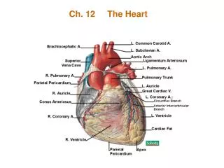
Ch. 12 The Heart
230 likes | 384 Vues
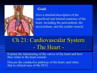
Ch. 12 The Heart. -The function of the heart is to pump blood through the arteries, veins, and capillaries. -The heart is located in the Thoracic Cavity, called the MEDIASTINUM -The Heart is enclosed in 3 Pericardial Membranes. --Fibrous (pericardial sac) --Parietal

Ch. 12 The Heart
E N D
Presentation Transcript
-The function of the heart is to pump blood through the arteries, veins, and capillaries. -The heart is located in the Thoracic Cavity, called the MEDIASTINUM -The Heart is enclosed in 3 Pericardial Membranes. --Fibrous (pericardial sac) --Parietal --Visceral (epicardium)
The heart has 4 chambers 1. Right Atrium 2. Left Atrium 3. Right Ventricle 4. Left Ventricle The chamber walls are made of cardiac muscle called Myocardium The Chambers are separated by valves, which also prevent the backflow of blood.
The Atria are the upper chambers The RIGHT ATRIUM - receives blood from the upper body - has a tricuspid valve(3 flaps) called the atrioventricular valve (AV) -the Superior Vena Cava brings blood from the upper body -The Inferior Vena Cava brings blood from the lower body.
The LEFT ATRIUM -receives blood from the lungs via the Pulmonary veins -has a bicuspid valve (2 flaps), called the mitral valve.
The Ventricles are the lower chambers RIGHT VENTRICLE -pumps blood to the lungs via the pulmonary artery. -has a semilunar valve between the ventricle and pulmonary artery.
LEFT VENTRICLE - pumps blood through the body via the AORTA (the largest artery) - has a Aortic Semilunar Valve between ventricle and aorta. - has thicker walls, which allow to contract forcefully. The Aorta is the largest artery in the body. Located on the top of the heart.
Coronary Vessels: - Purpose is to supply blood to the Myocardium (heart muscle) - The path it follows is: Aorta, to large arteries, to smaller arteries, to arterioles, to capillaries, to veins, and back to the right atrium. - An obstruction causes the mycardium to lose its blood supply. This is called ISCHEMIA.
-Prolonged Ischemia results in an INFARCT, or area of dead (necrotic) heart tissue. - Myocardial Infarction is a heart attack
The Cardiac Cycle: is a sequence of events in one heartbeat. It keeps the blood moving through the body. -The contraction is called SYSTOLE -The relaxation is called DIASTOLE -Atrial systole is followed by ventricular systole. When atria are in diastole, the ventricles are in systole.
The sound the heart makes is Lub-dub • -Lub is caused by ventricular systole closing the AV valve. This is the loudest and longest sound heard. • -Dup is caused by the closure of the semilunar valves • Heart murmurs are extra sounds that can be heard. • From valves that are too small (Stenosis) • Valves not closing properly
Cardiac Conduction Pathway - is the electrical activity of the heart - SA node (Sinoatrial) is the natural pacemaker of the heart. --The SA node sends impulses to the Atrioventricular (AV) node. - The AV node then delivers the impulse to the atrial myocardium, and then to the Bundle of His.
ECG stands for electrocardiogram • -this is the measure of the heart’s electrical activity. • * Atrial Activity is the P waves • * Ventricular Activity is the QRST wave • Arrhythmias are irregular heartbeats • Palpitations are an awareness of irregular beats • Fibrillation is a very rapid and uncoordinated ventricular beat.
Bradycardia is having a rate less than 60 beats/min. With an exception for athletes. Tachycardia is have a rate over 100. * The Heart Rate is regulated by the Nervous system.
Heart Diseases: - Atherosclerosis: cholesterol is deposited on the artery walls, causing a narrowing of the arteries, and a decrease in blood flow. Can also result in a formation of a clot. - hypertension: high blood pressure causes the left ventricle to work harder, enlarging it and making it weak.
Flow of blood through the body is: Superior/Inferior Vena Cavas Right Atrium A.V. Valve (tricuspid) Right Ventricle Pulmonary Semilunar valve Pulmonary Artery Lungs Pulmonary Vein Left Atrium Mitral Valve (bicuspid) Left Ventricle Aortic Semilunar Valve Aorta Body
-The right side of the heart deals with Deoxygenated Blood (no oxygen) -The left side of the heart is the Oxygenated blood (with oxygen) -Veins return blood to the heart, Arteries take blood from the heart.
Heart Rate = Pulse -Adult Resting Pulse = 60-80 beats/min. -Childs normal pulse = up to 100 beats/min -Infants normal pulse = as high as 120 beats/min. -Near Term Fetus = as high as 140 beats/min. ** The smaller the individual, the greater the pulse.