Download
1 / 21
320 likes | 1.96k Vues
Rhabdoviridae. PROPERTIES Bullet shaped, enveloped virion with helical nucleocapsid Genome is negative sense ss RNA Large glycoprotein peplomeres – G protein Neutralizing antibodies are directed against G protein epitopes Cytoplasmic replication – virions bud from cytoplasmic membrane

E N D
Rhabdoviridae • PROPERTIES • Bullet shaped, enveloped virion with helical nucleocapsid • Genome is negative sense ss RNA • Large glycoprotein peplomeres – G protein • Neutralizing antibodies are directed against G protein epitopes • Cytoplasmic replication – virions bud from cytoplasmic membrane • Some rhabdoviruses cause RAPID CYTOPATHOLOGY – e.g. vesicular stomatitis virus, others are NONCYTOPATHIC – e.g. rabies • During replication, defective interfering particles – DI – are commonly formed. DI particles are deletion mutants with greatly truncated genome – T particles – that interfere with replication of normal infectious virions
Rhabdoviridae • Rhabdoviruses are thermolabile, sensitive to UV light, and are readily inactivated by detergent-based disinfectants • e.g. QUATS –Zephiran, Cidex and Nolvasan • Genera • Genus Lyssavirus – Rabies virus • Genus Vesiculovirus – Vesicular stomatitis virus • Genus Ephemerovirus – Bovine ephemeral fever virus • Genus Novirhabdovirus – Infectious hematopoietic necrosis virus of fish
Rhabdoviridae • Genus Lyssavirus – Gr. Frenzy • Genus contains rabies virus and rabies like viruses – MOKOLA virus • Lagos bat virus • Duvenhage virus • European bat viruses 1 and 2 • Australian bat lyssavirus • BATS are the reservoir hosts • Lyssaviruses are relatively noncytopathogenic, thus encephalitis and death occur in many cases of infection with little or no cell destruction
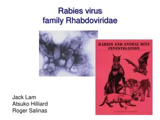
Rhabdoviridae - Rabies • Rabies – latin “rage” • One of the oldest and most feared disease of humans and animals, most lethal of all infectious diseases • Distribution –worldwide • Rabies free – • Japan, New Zealand, hawaii, Antartica, some of Europe, Great Britian, most Caribbean
Rabies • Australia – Australian bat lyssavirus • NA and Europe, wildlife threat is increasing • Worldwide – dog rabies is the cause of 40,000-50,000 human cases/year • Cattle rabies is a problem in Central and SA
Rabies • Hosts – all warmblodded animals have variable degrees of susceptibility • Foxes, coyotes, jackals, wolves and certain rodents are among the most susceptible animal groups • Skunks, bats, raccoons, rabbits, cattle, some members of the Felidae family and Viverridea – civet, mongoose etc. High susceptibility
Rabies virus • Etiologic agent – lyssavirus • In north america – six lyssavirus strains have been identified • Two skunk strains, an arctic fox and red fox strain, gray fox s train, dog/coyote strain and a racoon strain • Virus extremely labile when exposed to UV light and heat • At 20 degrees C – virus cannot survive for more than 25 hours in carcasses
Transmission – bite, scratch of rabid animal with the virus in its saliva; Aerosol transmission in cave dwelling bats; Human rabies from corneal transplant • Skunks – most important for perpetuation of wildlife rabies problem. They account for most causes of cattle rabies • High prevalence • Excretion of large quantities of virus in their saliva during the prolonged period 4-18 days of clinical disease
Rhabdoviridae - Bats • Bats – ability based on protracted clinial course not a subclinical carrier state • Subclinical infection can progress to more advanced clinical disease during times of stress. • Survival rate is high in bats • Cases of rabies in insectivorous vats • Vampire bats – major roel in human and animal rabies in Mexico, Central america, South America • Latin america – 250-500,000 cases per annum–vampire bats • Australian bat lyssavirus – isoltated from fruit eating bat in Queensland- two humans died
Rhabdoviridae • Pathogenesis – • infection in muscle until adequate amounts reach motor or sensory nerve endings in the muscle or skin • Virus shed from myocytes into extracellular spaces, bind to Ach receptors – other receptors facilitating its entry into nerve endings • 2nd phase of infection begins when the virus progresses centripetally to the CNS via the axoplasm of the peripheral and central nerves • The virus reaches the limbic system where it replicates extensively, leading to the furious form of rabies. Spread within the CNS continues with replication in the neocortex, resulting in the dumb or paralytic form • Late in infection, virus moves centrifugally from the CNS down the peripheral nerves to a variety of organs including the adrenal cortex, pancreas and the salivary glands via cranial nerves • Extensive replication in the salivary glands results inHIGH CONCENTRATIONS OF VIRUS IN THE SALIVA
Rhabdoviridae – Pathology and Immunology • Pathology - • Histopathologic examination of the brain reveals that many neurons are infected. • No frank cytopathology • little inflammatory cell infiltration • minimal target damage • Lethal neurologic dysfunction • Immunology – Humoral and cell-mediated immune responses cannot be detected during the time the virus moves from the site to the bite to the CNS • Infection is noncytopathic in muscle and nerve cells • Very little viral antigen is release to stimulate host defense mechanisms • NEURONS DON’T EXPRESS MHC CLASS I PROTEINS
Rabies– Clinical Features • IP – 14-90 days, but may be longer • IP is influenced by dose, strain of virus, degree of innervation and site of inocultion • Ascension in nerves is 8-16 mm per day • Prodromal period – period of virus shedding and change in temperament before obvious clinical disease is observed • 2 clinical forms – initial furious form and a terminal dumb form • Death results from respiratory failure • A higher proportion of cats and horses exhibit fury than is the case for cattle or other ruminants or laboratory animals • In dogs or cats, rare individuals survive more than 10 days after virus is first shed into the environment
Diagnosis – Direct FAT – demonstrate rabies antigen in touch impressions of brain tissue (medulla, cerebellum, hippocampus) Reverse transcription-polymerase chain reaction RT-PCR to test for the presence of viral RNA in the brain of the suspect animal Antemortem diagnosis – humans only FAT, RT-PCR is performed using a skin biopsy, skin bx, corneal impression, saliva Negri bodies in the hypothalamus, thalamus, pons, cerebral cortex and dorsal horns of the spinal cord Not all virus positive brains show Negri bodies 75% in humans, lower in some wildlife species Virus isolation by intracerebral inoculation of weanling mice with fresh homogenized tissue. Control mice are inoculated with extracted tissue incubated with specific neutralizing antibody. Mice develop encephalitis within 14 days Rabies Diagnosis
Rhabdoviridae- Rabies control • Rabies free countries – quarantine involving segregation of dogs and cats in licensed premises for 6 months • Endemic countries – Vaccination of dogs and cats – wildlife vaccination • Post-exposure treatment in humans • Wash wound thoroughly with aqueous soap solution or QUAT • Human rabies immune globulin HRIG is used at 20 IU/kg of body weight – half infiltrated around the wound, half injected intramuscularly • US – Two human rabies vaccines are licensed – • Human diploid cell-culture vaccine – HDCV and rhesus kidney cell-culture adjuvanted vaccine – RVA • Protocol consists of five 1 mL doses given IM in the deltoid area, one each on days 0,3,7,14 and 28 • If a person was vaccinated previously, NO HRIG IS GIVEN, instead, two doses of vaccine are given one each on days 0 and 3
Vesicular stomatitis – VS – sporadic disease of cattle and other ruminants, horses, swine and humans Characterized by fever and vesicle formation Found in the Americas and Caribbean Etiologic agent – Vesiculovirus – 3 serotypes and several subtypes exist NO CROSS PROTECTION AMONG SEROTYPES Indiana serotype, New Jersey serotype, Isfahan serotype – all 3 subtypes. NJ serotype is the most common and has the widest distribution Virus can remain stable for days or weeks in cool water, soil and on vegetation It is inactivated in 10 minutes by 1% formalin, Roccal (QUAT)etc Genus – Vesiculovirus Vesicular Stomatitis
Genus Vesicular Stomatitis • Transmission – contact with contaminated milking machines – teat and udder lesions • Ingestion of contaminated fomites – mouth lesions • Arthropods – though the virus replicates in black flies, sandflies, leaf hoppers, culicoides and mosquitoes, mechanical transmission is believed to be more significant than biological transmission • Houseflies are mechanical vectors • Pathogenesis – enters the body through breaks in the mucosa and skin or by the bites of flying arthropods • Vesicle formation is a result of cell destruction, interstitial edema that separates the epithelium from underlying tissues • Spread of lesions occurs by local extension. Systemic and viremic phase of infection is observed only in pigs and small lab animals
Vesicular Stomatitis • Clinical Features – • IP 1-5 days • Vesicular lesions on the tongue, lips, gums, teats and coronary bands • Oral lesions are accompanied by profuse salivation and anorexia • Uncomplicated cases fully recover in 2 weeks • Diagnosis – • Virus isolation from vesicular fluids and tissue scrapings • Cell cultures, embryonated eggs and sucking mice – IC inoculation • Paired acute and convalescent serum samples – Antibodies are quantitated using CFT and VN test
Vesicular Stomatitis - Vaccine • Prevention and Control – • Vesicular stomatitis is a reportable disease • Quarantine of affected premises. • Movement of cattle, swine, and horses from affected premises is prohibition until 30 days after the last clinical signs or the disease is evident – with the exception of animals going directly to slaughter • Vaccination – USDA approved autogenous killed vaccines are available only during outbreaks – efficacy is in doubt • Horses – no lab tests to differentiate between a positive serological reaction dueto vaccination or natural exposure • The American Horse Council and AAEP recommend that horse owners and vets carefully consider the repurcussions from using vaccines – some states and countries require NEGATIVE BLOOD TESTS FOR ENTRY • Zoonotic – Humans – flu like symptoms for one week
Genus EphemerovirusBovine Ephemeral Fever – 3 d. sickness • BEF is a benign • noncontagious arthropod-transmitted • Acute febrile disease of cattle and water buffalo • Distribution – Africa, Asia and Australia – Never reported in N.A. • Etiologic agent – Bovine ephemerovirus – 1 serotype • Transmission – insect vectors have not been specifically identified but are believed to be Culicoides spp. And possibly mosquitoes
Bovine ephemeral fever • Pathogenesis – poorly understood • Virus is associated with the buffy coat fraction of the blood • Site of replication is unknown • Lesions include a serofibrinous polyserositis affecting the joints, pleural and peritoneal surfaces. Some lung edema and atelectasis • Effusion fluids are characterized by the presence of large numbers of neuts. The neutrophil influx, combined with evidence of increased permeability of small serosal vessels, suggests that BEF may represent an acute, neutrophil-dependent, immune complex hypersensitivity • Clinical Features – IP 2-4 days, Morbidity 80%, Mortality 1-2% • Sudden onset, inappetance, pyrexia, lameness • Severe drop in milk production in lactating cows • Excessive salivation, lacrimation, serous nasal discharge, drooling, and dyspnea • Abortion is observed in 5% of pregnant cows • Recovery is complete in 3 days (2-5), some remain recumbent for long periods after the temperature returns to normal
Bovine Ephemeral Fever- Vaccine • Diagnosis – • Virus isolation is difficult but can be attempted by inoculation of infected buffy coat cells into cell cultures derived from Aedes albopictus or IC inoculation of suckling mice • FA staining of blood smears prepared during viremia • Paired serum samples – ELISA or VN tests • Control – Vector control • Attenuated vaccines are used in endemic areas • Recovering animals have long-lasting immunity