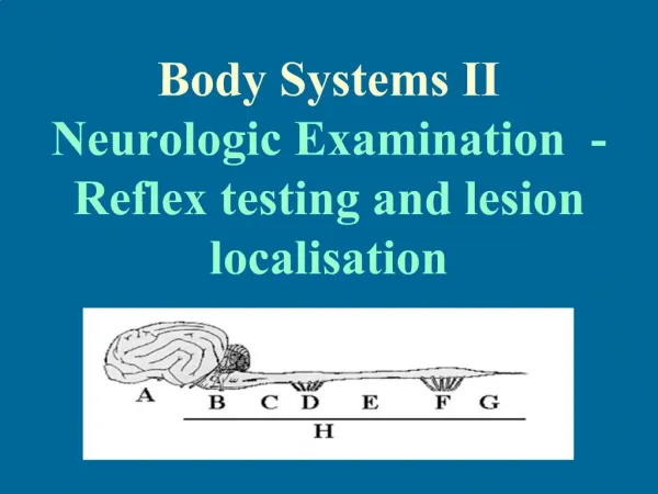
The Neurologic Examination
The Neurologic Examination. Debbie King FNP, PNP Fall 2010. Introduction. Purpose:: To present an organized technique for a complete neurological exam To make the exam less intimidating. History: Primary importance with neurologic disease. History Targeted topics Seizures Pain

The Neurologic Examination
E N D
Presentation Transcript
The Neurologic Examination Debbie King FNP, PNP Fall 2010
Introduction • Purpose:: To present an organized technique for a complete neurological exam • To make the exam less intimidating
History:Primary importance with neurologic disease • History • Targeted topics • Seizures • Pain • Gait coordination • Weakness • Tremor • History and ROS- may include • Subtle findings. • Transient findings. • Evaluations that require imaging.
Health History • Common or concerning symptoms- discovered in the review of systems • Headache • Dizziness or vertigo • Generalized, proximal, or distal weakness • Numbness, abnormal or loss of sensations • Loss of consciousness, syncope, or near syncope • Tremors of involuntary movements
Perform a complete, general examination • Full physical exam • May be patient’s only encounter with health professional. • Include all systems.
Central and Peripheral Nervous System — Key Definitions The Brain 4 regions: cerebrum, diencephalon, brainstem, cerebellum Contains interconnecting neurons (cell bodies and axons) Gray matter: aggregations of neuronal cell bodies White matter: neuronal axons coated with myelin Central nervous system: the brain and spinal cord
Brain and Spinal Cord • Interrelationship of the nervous system • Allows the body to perform the following: • Receive sensory stimuli • Identify and integrate the adaptive processes to maintain current body function • Orchestrate body function changes for survival • Integrate the rapid responsiveness of the CNS with the slower endocrine system • Control subconscious and involuntary body functions
The Brain and Spinal Cord • Protected by: • Skull • Vertebrae • Meninges • Cerebrospinal fluid • Circulates between an interconnecting system of ventricles in the brain and around the brain and spinal cord (shock absorber)
The Brain • Four regions • Cerebrum • Cerebellum • Brainstem • Diencephalon located here • the cranial nerves III-XII arise • FYI: Cranial nerves I and II emerge from the fiber tracts of the brain
Cerebrum • Two cerebral hemispheres • Each divided into lobes • Gray outer layer, the cerebral cortex • Higher mental function • General movement • Visceral function • Perception • Behavior • Integration of all the above
Cerebrum • Lobes • Frontal • Motor cortex for voluntary skeletal movements • Fine repetitive motor movement • Control of eye movement • Specific areas in the primary motor area-associated with specific parts of the body • Corticospinal tracts extend from the primary motor area in the spinal cord
Cerebrum • Parietal • Responsible for processing sensory data • Assists with interpretation of tactile sensations • Visual • Gustatory • Olfactory • Auditory sensation • Awareness of body position (proprioception) • Fibers communicate between the sensory and motor areas of the brain
Cerebrum • Temporal lobe • Responsible for perception and interpretation of sounds and determination of tier source • Contains the Wernicke speech area • Permits comprehension of spoken and written language • Integration of behavior, emotion and personality • Long term memory is associated here
Cerebrum • Limbic system • Mediates certain patterns of behavior that determine survival • Reactions to emotions such as anger, love • Expression of affect is mediated by connections between the limbic system and the frontal lobe
Cerebellum • Aids the motor cortex of the cerebrum • Integration of voluntary movement • Processes sensory information from: • Eyes • Ears • Touch receptors • Musculoskeletal • Integrated with vestibular system • Uses the sensory data for reflexive control
Brainstem • Pathway between the cerebral cortex and spinal cord • Controls many involuntary functions • Structures include • Medulla oblongata • Pons • Midbrain • Diencephalon
The spinal cord Extends from brainstem (medulla) to L1-L2 vertebrae Contains motor and sensory pathways that exit and enter the cord via anterior and posterior nerve roots and spinal and peripheral nerves 5 segments: cervical (C1-8), thoracic (T1-12), lumbar (L1-5), sacral (S1-5), coccygeal Central Nervous System – Brain and Spinal Cord Note: Cauda equina at L1-2, where nerve roots fan out like a horse’s tail
Peripheral Nervous System — Motor and Sensory Pathways and Dermatomes • Motor and sensory pathways: descending motor and ascending sensory pathways • Dermatome: band of skin innervated by the sensory root of a single spinal nerve
Spinal Nerves-FYI • Spinal nerves have motor fibers and sensory fibers. The motor fibers innervate certain muscles, while the sensory fibers innervate certain areas of skin. A skin area innervated by the sensory fibers of a single nerve root is known as a dermatome. A group ofmuscles primarily innervated by the motor fibers of a single nerve root is known as a myotome. Although slight variations do exist, dermatome and myotome patterns of distribution are relatively consistent from person to person
Spinal Nerves- both Dermatomes and Myotomes • Cervical plexus, C1 - C4, innervates the diaphragm, shoulder and neck • Brachial plexus, C5 - T1, innervates the upper limbs • Lumbar plexus, T12/L1 - L4, innervates the thigh • Sacral plexus, L4 - S4, innervates the leg and foot
Physical Exam- Neurological • Incorporate all components in an organized exam • Initial Impression • Mental Status • Cranial Nerves • Motor • Deep Tendon Reflexes • Sensory • Cerebellar • Gait
Physical Exam Areas To Examine • Initial Impression • Gait, expressions, general habitus, body type, involuntary movements, aides, eyes, hands/skin, speech, presence of family members.
Gait • Shuffling • Ataxic • Hemiparetic • Antalgic limp
Expressions • Masked facies (Parkinson’s) • Indifferent • Nervous • Apprehensive • Hostile
General Habitus • Clean • Disheveled • Dirty • Smelly
Body Type • Cushingoid • Acromegalic • Hirsute • Alopecia (male pattern)
Involuntary Movements • Tics • Myoclonus • Petit mal seizure • Tremor
Aides • Hearing • Walker • Cane • Wheelchair
Eyes • Exophthalmos (uni/bilateral) • Ptosis • Pupillary asymmetry
Presence of Family Members • How is the patient interaction? • Who does the talking? • Are there any pediatrics, geriatric, dependency, dominance issues.
Mental Status Exam at a glancechapter 5 in text • Physical appearance and behavior • Grooming • Emotional status-mood • Body language • Thoughts and perceptions • Cognition • Memory • Attention • Information • Vocabulary • Calculations • Abstract thinking • Constructional ability
Mental Status • Level of consciousness immediately assessed (grossly) • Neurological and psych findings are frequently coincidental • Depression and anxiety may be manifested in cc. • Establish if the patient is uncooperative vs. unable to understand.
Mental Status-Emotional State • Goal-Differentiate subtle changes vs. emotion vs. global findings. • Appropriate/inappropriate and how • Depressed, Angry, Ambivalent
Mental Status- Exams • Perform Mini Mental State • 11questions • Takes about 5-10 minutes • Examples of questions; • Year/season/date/day/month • Show five items, in 10 minutes ask what the items were • http://utswfm.googlepages.com/NH_MMSE.pdf • Cognitive Impairment Test • Correlates well with the MMSE • Six question • Performs better to detect milder forms of dementia • http://www.patient.co.uk/doctor/Six-Item-Cognitive-Impairment-Test.htm
Mental Status- Recall • Recent Memory • Name three unrelated items • Repeat immediately • Repeat in 5 minutes • Barn, computer, car, etc. • Testing for aphasia • Word comprehension • Repetition • Naming • Reading comprehension • Writing • Found in Chapter 5 of text
Mental Status-Memory • Remote or distant memory • Ask about verifiable past events • Mothers maiden name • Name of high school • Intermediate memory • Current events
Mental Status-Attention & Calculation • Attention Span • Follow a series of short commands • Repeat a story • Spell a word backwards • Calculation • Serial 7: 100-7 • Any arithmetic calculation
Mental Status-Language • Name-Common item e.g., watch, pen • Repeat “no ifs ands or buts”, “Methodist Episcopal” • Follow 3 step command-touch index finger of left hand to nose and ear. • Read “close your eyes” and follow commands.
Mental Status-Lang. Cont’d • Write a sentence • micrographia in Parkinson’s • Omission or addition of letters, words or mirror writing may indicate aphasia • Copy a design • Uncoordinated writing or drawing may indicate dementia, parietal lobe damage, a cerebellar lesion, or peripheral neurophathy