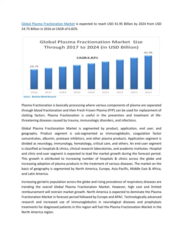
Sub-Cellular Fractionation
Sub-Cellular Fractionation. The experiment focusses on the separation of different organelles like chloroplast, mitochondria, golgi bodies so as to analyse their proteome content separately from a single sample. Related LOs: Cell composition, Culturing of cells

Sub-Cellular Fractionation
E N D
Presentation Transcript
Sub-Cellular Fractionation The experiment focusses on the separation of different organelles like chloroplast, mitochondria, golgi bodies so as to analyse their proteome content separately from a single sample • Related LOs: Cell composition, Culturing of cells > Prior Viewing – IDD-1. Extraction of bacterial protein, IDD-6. Extraction of serum protein. > Future Viewing – IDD-11. Protein quantification, IDD-14. Isoelectric focusing, IDD-17. SDS-PAGE • Course Name: Sub-Cellular Fractionation • Level(UG/PG): UG • Author(s): Dinesh Raghu, Vinayak Pachapur • Mentor: Dr. Sanjeeva Srivastava *The contents in this ppt are licensed under Creative Commons Attribution-NonCommercial-ShareAlike 2.5 India license
Learning objectives 1 After interacting with this learning object, the learner will be able to: • Define the sucrose density gradients to carry out organelle fractionation. • Operate the steps to retrieve the cells from the culture flask. • Practise to lyse and separate the organelles from the cell. • Infer the steps involved to perform experiemnt. • Assess the troubleshooting steps involved in the experiments. 2 3 4 5
Master Layout 1 Sample preparation (Slide: 5-9) Buffertreatment (Slide: 10) 2 Cell homogenization (Slide: 11-12) 3 Reagent Preparation(Sucrose Solution) (Slide: 13-14) Preparation of Density gradient column (Slide: 15-16) 4 Retrieving the organelle (Slide: 17-20) 5 Animate for user click to show images or instruments used for each step from the respective slides.
Definitions and Keywords 1 1. Subcellular Fractionation: The process by the cell organelles are separated on the basis of density of the organelles by the density gradient column . 2. Sucrose density gradient centrifugation: The process or condition at which the organelles get separated when the density of the organelles equal to the density of sucrose (Disaccharide ,carbohydrate) in the column. 2 3 4 5
Step 1: T1: sample preparation 1 2 3 cells 4 Incubator Culture flask Box with ice 5
Audio Narration Description of the action Step 1: T1: sample preparation 1 Cells are grown in the culture flasks at required set temperature in the CO2 incubator. The culture flask aids the cell growth by providing nutrients, surface area for the growth and cell division. Cells need to be taken out for sample preparation. Animator should draw a box containing ice. Draw a instrument labeled as”CO2 incubator” Animate like the user taking the culture flask (containing red solution) by opening the CO2 incubator as shown in figure. Make small round like structures and label it as “cells” inside the flask. Zoom the flask to show cells inside the culture flask. Animate like the user placing the culture flask on the ice. 2 3 4 5
Description of the action Audio Narration Step 1: T1: sample preparation 1 Cell scrapper 2 Cell scrapper action on culture flask 3 Animator should draw the cell scrapper as shown in figure. Animate like the user taking the cell scrapper, opening the culture flask containing red solution and the user should scrap the flask walls like the movement as shown in figure. Animate like the user taking the red solution from culture flask to the falcon tube as in figure Remove the adherent cells slowly by using the cell scrapper. The sample must be transferred to falcon tube. 4 5
Step 1: T1: sample preparation 1 2 3 4 rotor Centrifuge 5
Description of the action Audio Narration Step 1: T1: sample preparation 1 Instruct user to carry out centrifugation step. The animator should draw a centrifuge as shown in the figure. Animate in such a way that user clicks on lid to open it and keep the tube inside the rotor. Animate user control for setting parameters to set 3000g, 5minutes and click “enter” and animate like closing the lid and click “start”. Show a clock running for 5minutes . After 5 minutes, user should Click on STOP, OPEN and remove the tube out. • Please include the buttons like enter, set, start, open in the centrifuge display. Perform a low speed centrifugation at 3000g for 5 minutes. Centrifuge helps to settle the cells. 2 3 4 5
Step 2: T2: Buffer treatment 1 falcon tube 2 3 Description of the action/ interactivity Audio Narration (if any) . • Zoom the falcon tube to show formation of pellet at bottom and liquid layer on top of it. Let user take the pipette and show like pipetting out the top layer, to discard it. To the pellet, instruct user to add buffer. • Show buffer bottle, instruct user to open the bottle and set the pipette to 400ul and show like pipetting out the buffer and pouring into the tube. • The user should click on the pipette for the action to be done. Kindly redraw the figures Wash the pellet with phosphate buffer of pH 7.4 thoroughly to remove the excess broth. For more wash follow the same centrifugation step and add buffer, till the excess broth and color of broth disappears. Once broth is removed completely, cell lysis need to be carried out. 4 5
Step 3: T3:cell homogenization 1 ON Button 2 3 Description of the action/ interactivity Audio Narration (if any) Show homogenizer, instruct user to pick the instrument and falcon tube. Show like keeping the falcon on the ice box containing ice Now dip the rod into the solution (in Falcon) as shown in the figure. Instruct the user to click “ON” the instrument. Animate the solution getting mixed in all direction in the tube. Kindly redraw the figures Homogenize the tissue so that the cell lyse and the contents are released. Initially begin with low speed, later increase the speed accordingly. 4 5
Audio Narration (if any) Description of the action Step 3: T3:Cell homogenization 1 Post nuclear supernatant 2 Nucleus, cell debris 3 After homogenization carry out centrifugation and later keep the tube on ice for 5min. Show centrifugation as in slide 9&10. Animate formation of two layers in the tube as shown in the figure. and animate like when the user clicks on the level in tube, user must get the composition details. Nucleus and cell debris settle down as a result of low speed centrifugation and organelles remain in the supernatant as shown. 4 5
Description of the action Audio Narration (if any) Step 4: T4: Reagent Preparation(Sucrose Solution) 1 Falcon tube 2 3 Show a measuring balance, instruct user to tare the balance, let user place paper on the balance. Display balance reading as “0.003g”. now instruct to click on “0”/TARE option, once user clicks the button display the reading to 0.000g. Tarring of balance is very important before weighing the reagents. 4 5
Description of the action Audio Narration Step 4 : T4: Reagent Preparation(Sucrose Solution) 1 Prepare 15%-35% range sucrose gradient for preparing density gradient centrifugation to carry out fractionation. Draw the weigh balance, a spatula, bottle containing Sucrose and the 6 falcon tubes labeled as 15%, 20%, 25%, 27.5%, 30% and 35%. Instruct user to weigh sucrose. Animate user control to click on the sucrose bottles to measure 7.5g,10g 12.5g,13.75g,15g and 17.5g and put that into the respective falcon tubes. Draw a bottle labeled as distilled water and measuring cylinder. Animate like the user pouring 50ml of water to the cylinder (zoom and show the measurement) and then to the weighed Sucrose in falcons and show like mixing them. Animate sucrose powder getting into the solution, when user shakes the falcon. 2 3 4 5
Description of the action Audio Narration Step 5: T5:Preparation of Density gradient column 1 2 Post nuclear supernatant 3 Animate like the user taking the sucrose from different tube and add it to required amount as shown in the figure. Add 0.25ml of 35%, 0.5ml of 30%, 0.75ml of 27.5%, 1ml of 25%, 1ml of 20% and 1ml of 15%. Once the gradient is formed, instruct user to add 0.5ml post nuclear supernatant on top of the gradient. The sucrose addition must be on sides, to allow sucrose run down the tube. Pour sucrose gradient to the centrifuge tube and form a gradient for organelle separation and pour the post nuclear supernatant at the top to carry out centrifugation. 4 5
Step 6: T5:Preparation of Density gradient column 1 2 3 4 rotor Centrifuge 5
Description of the action Audio Narration Step 6: T6: Retrieving the organelle 1 • The animator should draw a centrifuge as shown in the figure. Animate in such a way that user clicks on open to open it and keep the tube inside the rotor (with lots of holes) as shown. The animator should animate user action to set the parameters for 132,000g , 16 hours and 30 minutes and click “enter” and animate like closing the lid and click “start”. Show a clock running for 16 hrs and 30 minutes . Once the time is done the user should open the lid by clicking “open” and remove the centrifuge out. • Please include the buttons like enter, set, start, open in the centrifuge. Transfer the content into the falcon tube and perform a low speed centrifugation at 132,000 g for 16hrs and 30 minutes at 4’ C. Organelles separate when the density of the organelle equals the density of the sucrose gradient 2 3 4 5
Step 7: T6: Retrieving the organelle 1 2 3 4 5
Description of the action Audio Narration Step 7: T6: Retrieving the organelle 1 Animate user taking out the tube from the centrifuge. Zoom the tube to show formation of different levels, color each of them differently. Now place the tube on a stand, with the help of needle make a opening at the bottom of the tube, to collect each fractions (levels) place a new tube at the bottom and label them accordingly after collection. animate the above action with user interaction like making holes and placing a fresh tube below the centrifuge tube. Please re-draw the figures from previous slide. Draw a freezer labeled as -80C and the user should keep the collected tube inside the freezer by opening the freezer Make a hole in the bottom of the tube and collect the sucrose content along with the separated organelles in the eppendorf tube and store at -80 C. care should be taken to collect each fraction separately in a fresh tube. the fractions can be cleaned and processed for further analysis. 2 3 4 5
Description of the action Audio Narration Step 7: T6: Retrieving the organelle 1 Animator should make animation as made in the SDS PAGE separation IDD as the next step of the experiment. The fraction samples collected can be used to run1D, SDS PAGE separation IDD as the next step of the experiment. • For more information and continuity follow the future viewing IDD like mentioned in slide:1. 2 3 4 5
Button 01 Button 02 Button 03 Slide 5-9 Slide 10 Slide 11-12 Slide 13-14 Slide 15-16 Slide 17-20 Tab 01 Tab 02 Tab 03 Tab 04 Tab 05 Tab 06 Tab 07 Name of the section/stage Animation area Interactivity area Instructions/ Working area Credits
Questionnaire: APPENDIX 1 Question 1 What is sub cellular components? a)Cell membrane b)Cell wall c)Channels D)Organelles and cytosol Question 2 What is subcellular fractionation? a)Separation of DNA of the cell b)Separation of different organelles c)Separation of RNA d)Separation of proteins
Questionnaire: APPENDIX 1 Question 3 What is Sucrose Density gradient? a)Column consisting of sucrose with uniform density b)Column consisting of sucrose with increasing density c) Column consisting of sucrose with uniform density equal to organelle density d) Column consisting of sucrose with uniform density equal to cell density Question 4: What is principle behind Sucrose Density gradient separation? a)Organelle separates when the density of sucrose is higher than organelle density b)Organelle separates when the density of sucrose is lower than organelle density c)Organelle separates when the density of sucrose equals organelle density d) Organelle separates when the density of organelle is higher than sucrose density
Questionnaire: APPENDIX 1 Question 5: Sucrose is a a)Monosaccharide ,Carbohydrate b)disaccharide, carbohydrate c)Polymer d)polysaccharide,carbohydrate
APPENDIX 2 Links for further reading Research papers: Huber LA,Pfaller K,Vietor I (2003). Organelle Proteomics:Implications of Subcellular Fractionation in Proteomics. Journal of american Heart Association.92:962-68. Mito Sciences (2007).Sucrose Gradient Separation protocol. BOOKS : Biochemistry by Stryer et al., 5th edition Biochemistry by A.L.Lehninger et al., 3rd edition Biochemistry by Voet & Voet, 3rd edition
APPENDIX 3 Summary The experiment was focused on the organelle separation by the density gradient column which works when the density of the organelle equal to the density of the sucrose gradient. Protocol for the crude sub-cellular fractionation of cultured mammalian cells that is both straightforward and cost effective and may facilitate the more accurate study of proteins and the generation of pure preparations of said proteins from cell extracts.