Download
1 / 53
530 likes | 1.63k Vues
Factors that affect Protein Activity and Protein Mechanism. Chapter 6 (Page 212-218, 225-231). 1. Factors that affect Enzyme Activity. Inhibitors Proteolytic activation Protein modifications Subunit cooperativity and modulators pH. 1I. Proteolytic Activation.

E N D
Factors that affect Protein Activity and Protein Mechanism Chapter 6 (Page 212-218, 225-231)
1. Factors that affect Enzyme Activity • Inhibitors • Proteolytic activation • Protein modifications • Subunit cooperativity and modulators • pH
1I. Proteolytic Activation In the ribosome, some proteins are synthesized as INACTIVE precursors. XXXX-E E + XXXX i.e. Trypsin begins as trypsinogen Protease Inactive Active
1I. Proteolytic Activation Nomenclature: Proprotein- For a protein precursor Proenzyme- Precursor for an enzyme Zymogen- Precursor for a protease
1II. Protein Modifications Some proteins following synthesis undergo covalent modifications other than cleavage, which affects their activity. • An example of this is the addition or removal of a phosphate group Protein Kinase Protein Phosphatase E-OH: Ser, Thr, Tyr
1II. Protein Modifications The covalent modification may affect catalysis. • By allowing the protein to adopt a conformation that may make it more or less active • By altering substrate-binding affinity • The binding of phosphate, a negatively charged molecule, could create electrostatic repulsion for a negatively charge substrate
1III. Subunit Cooperativity and Modulators Several enzymes are multimeric and their subunits engage in cooperativity (similar to hemoglobin binding of O2) in terms of their catalytic function. • These enzymes are generally called allosteric enzymes because they function through reversible, noncovalent binding of regulatory compounds called allosteric modulators.
1III. Subunit Cooperativity and Modulators Sigmoidal kinetic behavior is indicative of subunit cooperativity. This behavior is similar to hemoglobin binding of O2.
1III. Subunit Cooperativity and Modulators The effect of positive (+) and negative (-) modulators. Vmax remains constant but K0.5 changes. Vmax changes but K0.5 does not change.
1IV. pH Effect Enzymes have an optimum pH (or pH range) at which their activity is maximal. Vmax @ pH 1.6 Vmax @ pH 7.8
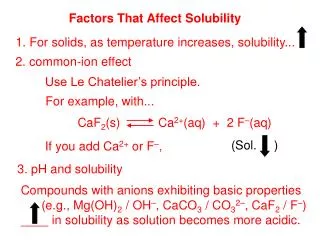
1IV. pH Effect • AA side chains in the active site may act as weak acids and bases with critical functions that depend on a specific ionization state. • AA ionization state is important for protein structural integrity. • The pH range over which an enzyme undergoes (greatest) changes in activity can provide a clue to the type of AA residue involved. • A change in activity near pH 7.0 could indicate the presence of an important His.
1IV. pH Effect • Recall that pKa values can shift based on the chemical environment.
2. Understanding Enzyme Mechanism Requires • Identification of all • Substrates • Cofactors • Products • Regulators (Inhibitors and activators)
2. Understanding Enzyme Mechanism B. Knowledge of • The temporal sequence of enzyme intermediate formations • The structure of each transition state • The structure of the enzyme intermediates • The rates of intermedateinterconversions • The energy contributed by all reacting and interacting groups, intermediate complexes, and transition states As of yet, the entirety of parameters involved in the mechanism of action is not known for any enzyme or protein in general.
The Mechanism of Action of Chymotrypsin
1. Chymotrypsin A 25 kDaproteolytic enzyme (serine protease) that catalyzes the cleavage of peptide bonds by a hydrolysis reaction. It cleaves proteins and peptides at the carboxyl terminal of a tryptophan, tyrosine, and phenylalanine AA residue.
2. Chymotrypsin Protein Structure His57, Asp102, and Ser195 form the catalytic triad.
3. Chymotrypsin Mechanism The free enzyme. pKa > 12.0 during the course of the reaction pKa significantly decreases
3. Chymotrypsin Mechanism The substrate. Stabilized in the oxyanion hole. Interacts with the hydrophobic pocket.
3. Chymotrypsin Mechanism Step 1: Substrate binding. When substrate binds, the side chain of the residue adjacent to the peptide bond to be cleaved nestles in a hydrophobic pocket on the enzyme, positioning the peptide bond for attack.
3. Chymotrypsin Mechanism Step 2: Nucleophilic Attack.
3. Chymotrypsin Mechanism Step 3: Substrate Cleavage. Instability of the negative charge on the substrate carbonyl oxygen leads to collapse of the tetrahedral intermediate; reformation of a double bond with carbon displaces the bond between carbon and the amino group of the peptide linkage, breaking the peptide bond. The amino leaving group is protonated by His57, facilitating its displacement.
3. Chymotrypsin Mechanism Step 4: Water comes in. An incoming water molecule is deprotonated by general base catalysis, generating a strongly nucleophilic hydroxide ion. Attack of hydroxide on the ester linkage of the acyl-enzyme generates a second tetrahedral intermediate, with oxygen in the oxyanion hole again taking on a negative charge.
3. Chymotrypsin Mechanism Step 5: Water attacks. Collapse of the tetrahedral forms the second product, a carboxylate anion, and displaces Ser195.
3. Chymotrypsin Mechanism Step 6: Break-off from the enzyme. Collapse of the tetrahedral intermediate forms the second product and displaces Ser195.
3. Chymotrypsin Mechanism Step 7: Product dissociates.
4. Evidence for Chymotrypsin Mechanism The rate dependence of chymotrypsin-catalyzed cleavage on pH shows a maximal velocity at nearly pH 8.0. The velocity was measured at low [substrate] so that
4. Evidence for Chymotrypsin Mechanism The turnover number (kcat) declines at low pH due to protonation of His57. The protonated His57 can not extract a proton from Ser195.
4. Evidence for Chymotrypsin Mechanism The changes in 1/Km reflect the deprotonation of Ile16, which engages in a salt bridge with Asp194. The loss of the salt bridge closes the hydrophobic pocket where the aromatic AA inserts. The substrate has a weaker affinity to the active site.
The Mechanism ofTransferrin Mediated Iron Delivery to Cells Courtesy of Anne B. Mason, Ph.D. University of VermontDepartment of Biochemistry
Iron Facts • Iron comprises 90% of the mass of the earth’s core • Iron is the second most common metal in the earth’s crust • Iron in its ferric form (Fe3+) is insoluble in water (10-18 M) • Most iron exists as ferric oxide hydrate=rust • Iron in its ferrous form (Fe2+) is extremely reactive via Fenton reactions
Borreliaburgdorferi Lactobacillus plantarum Posey & GherardiniScience288:1651-1653 (2000) Iron Facts All but two organisms require iron for life. It is important to maintain Fe homeostasis, a balance of the levels of Fe in the body.
The Problem with Free Fe in the Body • Free Fe refers to Fe not bound by a biomolecule. Fe3+ is the dominant form of Fe in our environment and though it is fairly harmless and mostly insoluble, it can be oxidized to Fe2+. • Fe2+ can generate reactive oxygen species. Uncontrolled levels of these species cause great harm to the body.
Iron-Bound Biomoleculesare Important to the Body • Metal binding by biomolecules maintains Fe in a bioavailable form. Many of these Fe-bound biomolecules play important roles. • Transport of small molecules • O2 and CO2 (as in hemoglobin) • Cofactor in important enzymes • Facilitates redox reactions
IronUptake Control completely at level of uptake • Heme (15-35% absorbed) • Red meat • Fish • Poultry • Non-heme (2-20%) • Lentils • Beans • Fortified cereals CDC
Iron Related Diseases • WHO considers iron deficiency to be the #1 nutritional disorder world-wide with ~80% of diets iron deficient • Iron related diseases • Sickle cell anemia • Thalassemia • Iron overload • Hemochromatosis (1 in 250 persons of Northern European descent) • Acquired iron overload (from multiple transfusions) • Iron implicated in a wide variety of other diseases, i.e., heart and liver disease, diabetes, neurodegenerative diseases, cancer and restless leg syndrome
Iron Containing Proteins Cytochromes RibonucleotideReductase Transferrin Ferritin Hemoglobin
Human Serum Transferrin N-lobe • Transferrin (TF) is an 80 kDa bilobal glycoprotein with 19 disulfide bonds • TF is synthesized in the liver and secreted into the plasma • Plasma concentration = 25-50 M • TF is 30% saturated with Fe • Knocking out TF in mice is lethal unless iron supplemented C-lobe Zuccola, HJ. PhD. Thesis (1992).
TF binds Two Fe(III) Ions • Ligand-to-metal charge transfer band (470 nm) in UV-vis produces characteristic pink color of TF HoloTF ApoTF • Binding AA groups come from two different domains in each lobe facilitating a change in conformation.
CII CI NI NII Fe(III) Binding Stabilizes TF “Open” “Closed” CII CI pH 5.6saltchelator Fe-chelator NI NII Apo Human TF Diferric Human TF (High Fe affinity) C ΔTm (HoloTF – apo TF) ~ 10 °
Transferrin Receptor (TFR) Transports Fe2TF into Cells dimer axes • TFR is a homodimeric protein found at cell membrane • Binds two molecules of Fe2TF • At pH 7.4 Fe2TF preferentially binds to the TFR (Kd is nM affinity) • At low pH (~5.6) apoTFpreferentially binds to the TFR (Kd is nMaffinity). Ectodomain (AA 89 - 760) Cytoplasmic region (AA1 - 67) Lawrence et al Science 286:779-782 (1999)
Low pH facilitates Fe release from TF N-lobe C-lobe Protonation of Lys296 and Lys206 in the N-lobe and of Lys534 and Arg632 in the C-lobe at low pH favors the “open” protein conformation and low Fe(III) binding.
Transferrin Mediated Iron Delivery to Cells Transferrin Mediated Iron Delivery to Cells ReceptorMediated Endocytosis Divalent Metal Transporter 1 Steere et al BiochemBiophysActa1820: 326-333 (2012)
Transferrin Mediated Iron Delivery to Cells Transferrin Mediated Iron Delivery to Cells ReceptorMediated Endocytosis • Two Fe2TF molecules bind to TFR. • The (Fe2TF)2-TFR complex is internalized into the cell via endocytosis. • In the endosome, a decrease of pH from 7.4 to 5.6 causes for a release of Fe(III) from TF. • The enzyme Steap3 reduces Fe(III) to Fe(II). • Fe(II) escapes the endosome via the divalent metal transporter I (DMTI) and is bound by other proteins for use in different ways.
Transport of Fe from Transferrin to Hemoglobin Fe2TF (Blood) • 75% of iron in the body is in hemoglobin • Acquisition of iron from transferrin is the rate limiting step in heme synthesis in developing red blood cells • Ferrochelatase is an enzyme bound to the inner membrane of the mitochondria • Ferrochelatase inserts Fe2+into protoporphyrin-IX forming heme. TFR Fe2TF-TFR Endosome & pH decrease Fe3+ Steap3 Fe2+ DMT1 & Ferrochelatase Hemoglobin
Paper Assignment on Enzymes