
Suturing
Suturing . Original by Rance Redhouse Lane Atene Kyle John Compiled by Craig Kohn Materials from Boston School of Medicine and other sources . Preparing the Wound . First, trim or shave the area surrounding the wound to avoid contamination and to ensure proper airflow.


Suturing
E N D
Presentation Transcript
Suturing Original by RanceRedhouse Lane Atene Kyle John Compiled by Craig Kohn Materials from Boston School of Medicine and other sources
Preparing the Wound • First, trim or shave the area surrounding the wound to avoid contamination and to ensure proper airflow. • The wound edges should be exposed and clearly visible. • Ideally, there should be a half-inch diameter of hair-free skin surrounding the wound.
Preparing the Wound (cont.) • Irrigating and washing the wound will remove bacteria and debris. Use soap to gently wash the skin wound and surrounding tissue. • Allow warm water to flow over and into the wound for a period of two full minutes. • This should be done immediately following the injury and three times daily until the wound is healed.
Preparing the Wound (cont.) • Following wound irrigation, pat the wound dry using a sterile gauze pad. • A clean paper towel can be utilized to dry the surrounding area. • Avoid using towels, as this can simply transfer additional bacteria to the clean wound.
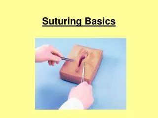
Supplies Materials Needed for Suturing
Curved Needle • Curved needles is probably the best needle you can use in the suturing process. • The curved needle are shaped like an arc to make the job easier and faster. • This needle can be used for any type of suture: continuous, non continuous, or purse string.
A needle holder is a surgical instrument used to hold a suturing needle for closing wounds during suturing and surgical procedures It has both a serrated portion and a cutting portion(for holding the needle and cutting the stitching material. Needle Holders
Needle Holding Techniques • There are several techniques for holding the needle holder. • The most common method is to place the thumb and ringfinger slightly into the instrument’s rings. • Avoid inserting your fingers far into the rings of the instrument, since this will tie up your fingers and impede your mobility. • Some surgeons do not put their fingers into the rings at all and simply grasp the rings and body of the needle holder in the palm of their hand. • Source: Boston University Medical Campus
Suture Materials • Suture materials can be divided into two categories: absorbable and non-absorbable. • Absorbable materials have the advantage in that they are less likely to cause an immune response by the body • Non-absorbable materials can be left in the body permanently if needed.
Absorbable Suture Materials • Absorbable suture materials are broken down by the patient’s body. • The original absorbable material was chromic catgut (still used today). • This is made from animal intestines and breaks down after 7 days. • Because it breaks down, there is less of a likelihood of an immune reaction.
Non-absorbable Suture Materials • Non-absorbable sutures are made of materials that are not readily broken down by the body’s enzymes or by hydrolysis. • Non-absorbable materials can be removed or left in place permanently • Source: Boston University Medical Campus
Forceps • Forceps allow you to control the position of the skin to make it easier to pass the needle and suture material through the skin. • For skin closure use a fine-toothed forceps • Source: Boston University Medical Campus
Skin hooks • Instead of using forceps, the skin edges can also be controlled using skin hooks • These have the advantage that they do not crush the skin edge.
Types of Sutures Continuous Sutures Non-continuous Sutures Purse-String Suture
Continuous Suture • A continuous suture, Also called uninterrupted sutureis made from an uninterrupted series of stitches that are fastened at each end by a knot. • A.k.a. Running Stitch
Non-Continuous Suture • Also called an Interrupted Stitch. • Each stitch is tied separately. • It be used in skin or underlying tissue layers. • This stitch has the benefit of creating a more accurate fit for the edges of the wound.
Pro’s and Con’s • Continuous + faster + less foreign material in wound + potentially better airtight/watertight - Knot failure, big deal - Less control over tension • Non-continuous + allows adjustment of tension + one knot failure, not a big deal - More time needed - Costs more - Increased amount of foreign material in wound • Courtesy of J. James
Purse-string Suture • A continuous stitch paralleling the edges of a circular wound. • The wound edges are inverted when tied. • Commonly used to close circular wounds, such as hernia or an appendiceal stump
Suturing Techniques Angles of Insertion Coordinating forceps and needle Key Maneuvers Knots Suture Removal
Remember to create right angles • The ideal skin suture should form a rectangle. • It should penetrate the epidermis and dermis perpendicular to the skin surface • After penetration, turn at a right angle, at the depth of the wound, move parallel to the skin surface, and then move straight to the surface.
Coordinated Use of the Forceps and Needle Holder • The tip of the needle holder should grasp the needle about 2/3 of the way back from the point. • The needle holder and needle should be perpendicularto each other. • The tip of the needle should penetrate the skin perpendicularly about 5-10 mm from the wound edge. • Elevate the skin with the forceps while penetrating the skin. 90*
Forceps & Needle (Cont) • The tip of the needle should now be seen protruding into the wound • At this point, continue to hold the skin w/ the forceps. • A common error here is to release the forceps from the skin edge • This would cause the skin to retract, and the needle may move and retract beneath the skin edge
Forceps and Needle (cont) • The key is to hold the position of the skin edge while releasing the needle from the needle holder. • Pull the needle from the other side of the elevated skin. • Elevate the other skin edge and penetrate it with the needle.
Knot Tying Principles • Simpler knots are better than complicated • Smaller knots are better than bigger • Excessive tension will cause tissue damage • Tension should be as horizontal as possible (minimize lifting) • Minimal ties per knot should be used; extra ties add bulk. • If the two ends of the suture are pulled in opposite directions with uniform rate and tension, the knot may be tied more securely. • Source: ruralareavet.org
Two-handed square knot • The two-hand square knot is the easiest and most reliable for tying most suture materials. • It may be used to tie surgical gut, virgin silk, surgical cotton, and surgical stainless steel. • Source: ruralareavet.org
Source: ruralareavet.org Two Handed Square Knot Steps
Surgeon’s Knot (Friction Knot) • The Surgeon’s Knot is recommended for synthetic materials or materials that do not easily hold their own shape. • The Surgeon’s Knot is similar to a Square Knot; however, the difference is that the first loop of the knot is double wrapped. • The left strand is wrapped twicearound the right strand. • The knot is finished just like a square knot
Suture Removal • Sutures should be removed from the… • Face: 3-4 days • Scalp: 5 days • Trunk: 7 days • Arm or leg: 7-10 days • Foot 10-14 days
Suture Removal Steps • The skin should be cleansed. • Hydrogen peroxide is a good choice for gently removing dried blood and exudate. • Grasp one of the “ears” of the suture with a forceps to elevate the suture just enough to slip the tip of a small scissor under the suture in order to cut it. • Source: Boston University Medical Campus
Suture Removal Steps • With the suture gently elevated, snip the suture with a scissors. • The suture is then gently removed by pulling with the forceps. • It is frequently a good idea to reinforce the wound with adhesive strips or tape to prevent it from re-opening.


![[READ DOWNLOAD] A NOVICE GUIDE TO SURGICAL SUTURING: Mastery Guide To Surgical Suturing](https://cdn7.slideserve.com/12525779/slide1-dt.jpg)