Syncope
Learning Objectives. Recognize the vast etiologies of syncopeUnderstand the importance of uncovering underlying organic heart diseaseLearn diagnostic and management strategies for neurally mediated syncope. You know, medicine is not an exact science, but we are learning all the time. Why, just fifty years ago, they thought a disease like your daughter's was caused by demonic possession or witchcraft. But nowadays we know that Isabelle is suffering from an imbalance of bodily humors, perhaps ca30040

Syncope
E N D
Presentation Transcript
1. Syncope Ed Da Veiga, M.D.
August 20, 2008
2. Learning Objectives Recognize the vast etiologies of syncope
Understand the importance of uncovering underlying organic heart disease
Learn diagnostic and management strategies for neurally mediated syncope
4. Case Presentation 38 year old male with hangover on flight for honeymoon to St. Lucia
Stewardess asks for medical assistance as patient �felt funny� and then �passed out�
What do you want to know?
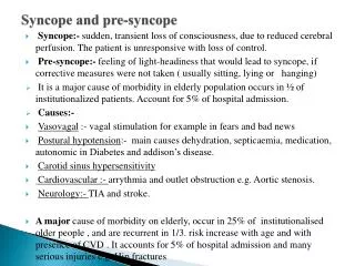
5. Overview Syncope is a symptom, not a disease
In all forms, consists of a sudden decrease or brief cessation of cerebral blood flow
Accounts for 3.5% of ER visits and 1-6% of all hospital admissions per year
6. Definition Sudden and brief loss of consciousness associated with a loss of postural tone, from which recovery is spontaneous
7. Distinguishing Syncope Dizziness, presyncope, and vertigo
No LOC or loss of postural tone
�Drop attacks�
Lead to falls without loss of consciousness
Sometimes sign of vertebrobasilar TIA (15%) Features to distinguish syncope from seizure
Prodromal/ Premonitory symptoms
Precipitating event
Events that follow it
8. Precipitants/Prodromal Symptoms LOC precipitated by pain, exercise, micturition, defecation, or stressful event usually syncope
Sweating, nausea = syncope
Aura = Seizure
Disorientation/ LOC > 5 minutes usually seizure rather than syncope
10. Important information WITNESSES?
Initial Assessment (especially HISTORY) will often lead to a clear diagnosis and help efficiently direct further workup and/ or treatment
H and P leads to identification of cause in 45% of patients
11. Differential Diagnoses Neurally Mediated Syncope (24%)
Vasovagal
Situational
Carotid Sinus
Orthostatic Hypotension (10%)
Psychiatric Disorders (2%)
Neurologic Dz (10%)
Cardiac Syncope
Organic Heart Disease (4%)
Arrhythmias (14%)
UNKNOWN (34%)
50-66% may be neurally mediated based on tilt-table studies
13. Structural Heart Disease Presence of a structural heart disease (CAD, CHF, Valvular Heart Disease, CHD) is the most important risk factor for predicting the risk of death
Have ? risk of death at one year
Most arrhythmias are found in these patients
15. Risk Factors Predictors of arrhythmic syncope or cardiac death at one year
CHF
Ventricular tachyarrhythmias
Abnormal ECG
Age >45 years
Presence of 2 or more of these is associated with >10% incidence of syncope or cardiac death
16. Cardiac Differential Cardiac Syncope: LOC often w/o prodrome
Indicates Outflow Obstruction
AS, HOCM, PAH, Pulmonic Stenosis, PE
MI, USA, Coronary Artery Spasm, Aortic Dissection
Arrhythmias
Prolonged QT (either Congenital or Drug Induced)
AV Block, Sinus Node Dysfunction
Ventricular tachycardia
Arrhythmogenic right ventricular dysplasia
Supraventricular tachycardia (Wolff-Parkinson-White)
17. Neurally Mediated Syncope Most Common Causes
Vasovagal, Situational, and Carotid Sinus Syncope
Results from sudden reflex mediated hypotension/ and or bradycardia
Triggered by various stretch/ mechanoreceptors (carotid sinus, bladder, esophagus, respiratory tract
19. Neurally Mediated Syncope Pathophysiology Peripheral Venous Pooling h causes sudden i in peripheral venous return
Leads to cardiac �hypercontractile� state which activates stretch receptors
Neural traffic h to brain mimics severe hypertension and provokes paradoxical bradycardia and i in PVR
20. TIMBER!!!
21. Orthostatic Hypotension Decline of >20mm Hg in SBP/ 10mm Hg in DBP from supine to standing
Supine HTN common in these patients
Elderly especially vulnerable
? Baroreceptor sensitivity, ? Cerebral Blood Flow, ? renal sodium wasting, ?thirst response with aging
Peripheral sympathetic tone impairment
Diabetic neuropathy, antihypertensive medication
22. Neurologic Causes Syncope rare manifestation of cerebrovascular disease
Subclavian steal syndrome,
Basilar Artery Migraine (syncope and HA)
Vertebrobasilar insufficiency �Drop Attacks�
23. Diagnostic Evaluation H and P! � 45% of time can identify cause
CBC, BMP
ECG- Low yield but can be important clues to look for underlying heart disease
CT Head, EEG: low yield
Echocardiogram/ Stress Test: Helpful when presence of underlying cardiac disease cannot be determined clinically
24. History Time of day
Activities preceding (recurrent/at rest, exercise associated, on standing)
Prodromes, associated symptoms
Duration of LOC
Injuries
Medications, ingestions
Cardiac History
26. Family History Sudden unexplained death
Deafness
Arrhythmias
Congenital heart disease
Seizures
Metabolic disorders
Myocardial infarction at young age
27. Physical Exam Pulse, blood pressure � taken supine and standing after 3 minutes
Murmurs, clicks of outflow tract obstruction
Neurologic examination
Carotid Massage (if no bruit)
28. Arrhythmia Testing Telemetry
Holter: 12-24 hours
symptoms w/ arrhythmia (5%) v. symptoms without arrhythmia (17%)
External Loop Recorders : can wear for weeks to months
Implantable Loop Recorders: Monitor for 12-18 months
Provided diagnosis in 55% of pts with unexplained syncope compared to conventional methods
EP Studies: Helpful with structural heart disease
29. Tilt Table Test Used to evaluate autonomic nervous system
Evaluates predisposition to neurally mediated syncope
Specificity of negative test 90%
30. Indications for Tilt Table Testing Unexplained recurrent syncope
Single episode associated with injury or in settings that pose a high risk of injury
If organic heart disease is present, than after cardiac causes have been excluded
Evaluation of recurrent syncope in setting of autonomic failure
Assessment of recurrent, unexplained falls
31. Indications for Hospital Admission History of CAD, CHF, Ventricular Arrhthmia
Accompanying Chest Pain
Abnormal ECG
Moderate to severe orthostatic hypotension
Age > 70 yrs
Resulting Trauma
32. Management
33. Management of Neurally Mediated Syncope
34. Patient Instructions Preventing Syncope or Vasovagal Spells
Avoid EtOH, lack of sleep, warm environment
Maintain adequate hydration and food intake
Avoid drugs that lead to hypotension
Avoid activities that precipitate syncope
Preventing LOC or Injury
Assume supine position upon onset of prodrome
Avoid driving or other activities that could lead to injury
35. Bibliography Kapoor, WN Syncope. NEJM 2000; 343: 1856-62
Freeman, R Neurogenic Orthostatic Hypotension NEJM 2008; 358: 615-624
Soteriades, et al. Incidence and Diagnosis of Syncope. NEJM 2002; 347:878-885
Grubb, B. Neurocardiogenic Syncope. NEJM 2005; 1004-1010
36. Thanks!