Download
1 / 44
440 likes | 461 Vues
Explore the process of apoptosis, or programmed cell death, and its importance in the development and health of organisms. Learn about the different triggers and stages of apoptosis, as well as the role of caspases and BCL-2 proteins. Discover how apoptosis is essential for proper cell function and the elimination of damaged or infected cells.

E N D
INTRODUCTION • Cell death by injury -Mechanical damage -Exposure to toxic chemicals • Cell death by suicide -Internal signals -External signals
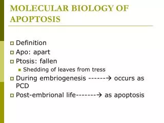
Conted….. • Apoptosis or programmed cell death, is carefully coordinated collapse of cell, protein degradation , DNA fragmentation followed by rapid engulfment of corpses by neighbouring cells. (Tommi, 2002) • Essential part of life for every multicellular organism from worms to humans. (Faddy et al.,1992) • Apoptosis plays a major role from embryonic development to senescence.
Why should a cell commit suicide? • Apoptosis is needed for proper development Examples: • The resorption of the tadpole tail • The formation of the fingers and toes of the fetus • The sloughing off of the inner lining of the uterus • The formation of the proper connections between neurons in the brain • Apoptosis is needed to destroy cells Examples: • Cells infected with viruses • Cells of the immune system • Cells with DNA damage • Cancer cells
What makes a cell decide to commit suicide? • Withdrawal of positive signals examples : • growth factors for neurons • Interleukin-2 (IL-2) • Receipt of negative signals examples : • increased levels of oxidants within the cell • damage to DNA by oxidants • death activators : • Tumor necrosis factor alpha (TNF-) • Lymphotoxin (TNF-β) • Fas ligand (FasL)
History of cell death / apoptosis research • 1800s Numerous observation of cell death • 1908 Mechnikov wins Nobel prize (phagocytosis) • 1930-40 Studies of metamorphosis • 1948-49 Cell death in chick limb & exploration of NGF • 1955 Beginning of studies of lysomes • 1964-66 Necrosis & PCD described • 1971 Term apoptosis coined • 1977 Cell death genes in C. elegans • 1980-82 DNA ladder observed & ced-3 identified • 1989-91 Apoptosis genes identified, including bcl-2, fas/apo1 & p53, ced-3 sequenced (Richerd et.al., 2001)
Necrosis vs. Apoptosis • Cellular condensation • Membranes remain intact • Requires ATP • Cell is phagocytosed, no tissue reaction • Ladder-like DNA fragmentation • In vivo, individual cells appear affected Necrosis Apoptosis • Cellular swelling • Membranes are broken • ATP is depleted • Cell lyses, eliciting an inflammatory reaction • DNA fragmentation is random, or smeared • In vivo, whole areas of the tissue are affected
NECROSIS Vs APOPTOSIS Wilde, 1999
STAGES OF APOPTOSIS Induction of apoptosis related genes, signal transduction Sherman et al., 1997
APOPTOSIS: Morphology organelle reduction membrane blebbing & changes cell shrinkage mitochondrial leakage nuclear fragmentation chromatin condensation Hacker., 2000
APOPTOSIS: Morphological events cell shrinkage organelle reduction mitochondrial leakage chromatin condensation nuclear fragmentation membrane blebbing & changes
Blebbing & Apoptotic bodies The control retained over the cell membrane & cytoskeleton allows intact pieces of the cell to separate for recognition & phagocytosis by MFs Bleb Apoptotic body MF MF
Caenorhabditis elegans 1090 cells 131 cells apoptosis decision to die engulfment degradation execution ced-3 ced-4 ced-1 ced-2 ced-5 ced-6 ced-7 ced-10 nuc-1 egl-1 ced-9 ces-2 ces-1
Death Receptors Initiator Caspase 8 Effector Caspase 3 PCD Initiator Caspase 9 Mitochondria/Cytochrome C Apoptosis: Pathways “Extrinsic Pathway” Death Ligands “Intrinsic Pathway” DNA damage & p53
Capsase activation • Comparison between active and inactive forms of caspases. Newly produced caspases are inactive. Specifically cleaved caspases will dimerize and become active.
The role of caspase • During apoptosis, the cell is killed by a class of proteases called caspases. More than 10 caspases have been identified. Some of them (e.g., caspase 8 and 10) are involved in the initiation of apoptosis, others (caspase 3, 6, and 7) execute the death order by destroying essential proteins in the cell. The apoptotic process can be summarized as follows: • Activation of initiating caspases by specific signals • Activation of executing caspases by the initiating caspases which can cleave inactive caspases at specific sites. • Degradation of essential cellular proteins by the executing caspases with their protease activity.
Introducing the BCL-2 family of proteins important in apoptotic pathways = anti-apoptotic Bcl-2 and related proteins part A
BH3 only protein binding specificity for BCL-2 homologues BIM and PUMA bind to all BCL-2 family members tested; by contrast NOXA only binds to A1 and MCL1. These binding specificities recapitulate the ability of these proteins to activate apoptosis e.g. BIM et al can induce apoptosis alone whereas a combination of NOXA and BAD is required. Youle and Strasser (2008) The BCL-2 protein family: opposing activities that mediate cell death. Nature Reviews Molecular Cell Biology, 9, 47-59
To summarize….. BCL-2 family of proteins have opposing apoptotic activities 1st set (e.g. Bcl-2 itself) inhibit apoptosis. 2nd set (e.g. BAX) promotes apoptosis. 3rd set (e.g. the BH3 only proteins) bind and regulate the anti-apoptotic BCL-2 proteins to promote apoptosis.
MAJOR PLAYERS IN APOPTOSIS • Caspases • Adaptor proteins • TNF & TNFR family • Bcl-2 family
Ligand-induced cell death Ligand Receptor FasL Fas (CD95) TNF TNF-R TRAIL DR4 (Trail-R)
Caspase • As shown in the above figure, a variety of death ligands (FasL/CD95L, TRAIL, APO-3L and TNF) can induce apoptosis. It is natural to see if they can kill cancer cells without affecting normal cells. TNF was first investigated in the 1980s for cancer therapy, but with disappointing results. Then CD95L (FasL) was tested in the 1990s. The results were still not satisfactory. Recently, TRAIL has been demonstrated to be highly selective for transformed cells, with minimal effects on normal cells. It could be an effective drug for both cancer and AIDS.
Death Domains Death Effectors Induced proximity of Caspase 8 Activation of Caspase 8 Ligand-induced cell death “The death receptors” Ligand-induced trimerization FasL Trail TNF
Death receptors Weinberg Fig 9.31 The extrinsic apoptotic pathway
APOPTOSIS: Signaling & Control pathways I Externally driven Externally driven Apoptotic signals Activators of initiator enzymes p53 Cytochrome C Internally driven Initiator caspases 6, 8, 9,12 mitochondrion Execution caspases 2, 3, 7 Apoptosis events Activation
APOPTOSIS: Signaling & Control pathways II Externally driven Externally driven Inhibitors Apoptotic signals Activators of initiator enzymes p53 Cytochrome C Internally driven Bcl2 Survival factors External Initiator caspases 6, 8, 9,12 Internal Execution caspases 2, 3, 7 Inhibitors of apoptosis Apoptosis events Inhibition
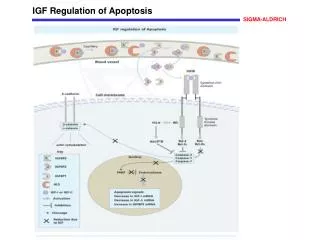
The mitochondrial pathway Growth factor receptors DNA damage Fas Casp8 PI3K p53 Akt Bid casp3 BAD Bid Bax IAPs casp9 Bid Apaf1 Bcl2 ATP casp3 Bax Cyt.C Smac/ DIABLO H2O2 AIF Pollack etal., 2001
REGULATION OF APOPTOSIS • Stimuli apoptosis selection of targets(Rich et al., 2000) • Apoptosis by conflicting signals that scramble the normal status of cell (Canlon & Raff, 1999) • Apoptotic stimulicytokines, death factors (FasL) (Tabibzadeh et al., 1999) • DNA breaks p53 is activated arrest cell cycle or activate self destruction(Blaint & Vousden, 2001)
Importance of Apoptosis • Important in normal physiology / development • Development: Immune systems maturation, Morphogenesis, Neural development • Adult: Immune privilege, DNA Damage and wound repair. • Excess apoptosis • Neurodegenerative diseases • Deficient apoptosis • Cancer • Autoimmunity
FUTURE PERSPECTIVES • The biological roles of newly identified death receptors and ligands need to be studied • Need to know whether defects in these ligands and receptors contribute to disease
CONCLUSION • an important process of cell death • can be initiated extrinsically through death ligands • (e.g. TRAIL, FasL) activating initiator caspase 8 through induced proximity. • can be initiated intrinsically through DNA damage (via cytochrome c) activating initiator caspase 9 through oligomerization. • Initiator caspases 8 and 9 cleave and activate • effector caspase 3, which leads to cell death.
DNA DAMAGE p53
The bcl-2 family BH4 BH3 BH1 BH2 TM N C Bcl-2 Group I bax Group II Bad bid Group III bik Receptor domain Raf-1 calcineurin Membrane anchor Ligand domain Pore formation phosphorylation Back
P53 & Apoptosis p53 first arrests cell growth between G1 S This allows for DNA repair during delay If the damage is too extensive then p53 induces gene activation leading to apoptosis (programmed cell death) BACK
3 mechanisms of caspase activation a. Proteolytic cleavage e.g. pro-caspase 3 b. Induced proximity, e.g. pro-caspase 8 c. Oligomerization, e.g. cyt c, Apaf-1 & caspase 9 Back
Apoptosis signal to kill infected cells Cytolytic lymphocyte/CTL (& natural killer lymphocyte) presents Fas ligand/CD178 on its surface totell the infected cell to die Fas ligand CTL Virally infected cell Externally driven Apoptotic signals Cytochrome c Initiator caspases Theimmunological synapse holds the cells much tighter together than shown here Execution caspases Apoptosis events Fas/ CD95 is the ‘death receptor’