Download
1 / 16
160 likes | 320 Vues
fMRI Activation of the Fusiform Gyrus and Amygdala to Cartoon Characters but not to Faces in a boy with Autism. Looking specifically at the fusiform gyrus during a fMRI differenciation task By Nidhi Joseph.

E N D
fMRI Activation of the Fusiform Gyrus and Amygdala to Cartoon Characters but not to Faces in a boy with Autism Looking specifically at the fusiform gyrus during a fMRI differenciation task By Nidhi Joseph
Autism is a neurodevelopmental disorder marked by social impairments, communication difficulties and stereotyped patterns of behaviour. Studies have shown that the right lateral fusiform gyrus (FG), an occipito-temporal visual cortical area, know as the fusiform facial area (FFA) is activated in humans when we look at faces; Gauthier et al., 1999 Objects of expertise are also found to recruit the FFA (Gauthier et al., 1999) So, specialization for faces in the FFA is an example of a more general phenomenon related to experience, individuation, and configural processing (Tarr & Gauthier, 2000) Objects activate the medial Fusiform Gyrus Introduction
Subjects • DD: • 12 year old boy with Autism • Special interested in Digimon, a Japenese cartoon series. Watched it daily • TDC: - A ‘typical’ 10 year old male • Aware of Digimon, but only watched it occasionally
Experimental task Subjects were shown unfamiliar faces and objects and Digimon characters. Two images at a time shown, from one category Asked to identify if they were the same or different using a button box
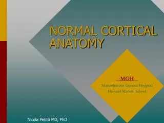
Tasks were performed while inside an fMRI scanner • 18 oblique coronal sections were viewed • The Green strip here, indicates the slides that the Fusiform Gyrus was located on
Results • DD: -no FFA activation to unfamiliar faces, activation to Digimon in the area where we would expect the FFA (t(30) = 6.38, P < 0.001) • TDC: -showed a greater response to unfamiliar faces than Digimon in the Fusiform Gyrus (t(31) = 3.44, P = 0.002) • INTERESTING! -Digimon with Masked faces- less activation in DD when compared up unmasked but more activation in TDC than to objects -TDC also showed least activation with familiar faces
MEAN PERCENT SIGNAL CHANGE DD TDC SUBJECT
Discussion • DD’s FG activation pattern- differences in the amount of time he has spent looking at or thinking about people and Digimon • Expert at Digimon (Gauthier et al., 1999) • Activation is possible even in the absence of prior “specialization” for faces • damage can be extremely selective (nearby areas can support non-face recognition like digimon) • Cannot say conclusively that the digimon faces aren’t involved in activating the FFA area • FFA decreases with repeated exposure to the same face (Buckner et al., 1998)
My opinion on the paper PROs • Visuals were great! • Congruent with other research • could be useful developing therapy for autism CONs • Slightly unclear • Brain images for TDC as well • Bigger sample size needed • Autistic people with expertise in different things, preferable non-face objects
References • Buckner, R. L., Goodman, J., Burock, M., Rotte, M., Koustaal, W., Schac-ter, D., et al. (1998). Functional-anatomic correlates of object priming in humans revealed by rapid presentation event-related fMRI. Neuron, 20, 285–296 • Gauthier, I., Tarr, M. J., Moylan, J., Skudlarski, P., Gore, J. C., & An- derson, A. W. (2000b). The fusiform “face area” is part of a network that processes faces at the individual level. Journal of Cognitive Neu- roscience, 12, 495–504, 912 • Grelottiet al., 2005. fMRI activation of the fusiform gyrus and amygdala to cartoon characters but not to faces in a boy with autism. Neuropsychologia 43(3):373-8 • Tarr, M. T., & Gauthier, I. (2000). FFA: A flexible fusiform area for subordinate-level visual processing automatized by expertise. Nature Neuroscience, 3, 764–769