Coronary Arteries and ECG Leads
1.33k likes | 1.47k Vues
Learn about the anatomy of coronary arteries and ECG leads to understand how the heart functions and how to interpret ECG readings for diagnosing cardiac conditions effectively.

Coronary Arteries and ECG Leads
E N D
Presentation Transcript
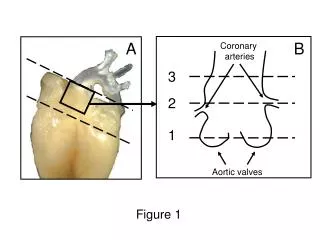
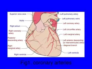
Coronary Arteries • Supply arterial blood to heart muscle • Left coronary artery carries about 85% of blood supply to myocardium • Right coronary artery carries remainder • Originate above aortic valve
Left Coronary Artery • Divides into left anterior descending and circumflex arteries • Left anterior descending (LAD) supplies: • Anterior wall of left ventricle • Interventricular septum • Circumflex supplies: • Lateral and posterior portions of left ventricle • Part of right ventricle
Coronary Arteries • Right coronary artery and left anterior descending artery supply: • Most of right atrium and ventricle • Inferior aspect of left ventricle • Anastomoses provide collateral circulation
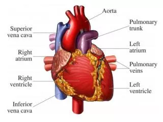
Coronary Capillaries • Exchange nutrients and metabolic wastes • Merge to form coronary veins • Coronary sinus empties into right atrium • Major vein draining myocardium
ECG Leads • Two surface electrodes of opposite polarity • Bipolar lead • Two electrodes of opposite polarity • Unipolar lead • Single positive electrode and reference point
Leads • Bipolar leads • Limb leads • I, II, III • Unipolar leads • Augmented limb leads • aVR, aVL, and aVF • Precordial leads • V1 through V6 • Each lead assesses electrical activity from a different angle
Standard Limb Leads • Record difference in electrical potential between left arm, right arm, and left leg electrodes • Represent axes
Axis • Average direction of the heart’s electrical activity • Triaxial reference system
Axis • Lead I is a lateral (leftward) lead • Assesses electrical activity from a viewpoint defined as 0° on a circle divided into an upper negative 180° and a lower positive 180°
Axis • Leads II and III are inferior leads • Assess the heart's electrical activity from vantage points of +60° and +120°
Bipolar Lead Placement Limb lead placement
Augmented Limb Leads • Same electrodes as limb leads • Record difference in electrical potential between extremity lead sites and a reference point • Zero electrical potential • At center of the heart’s electrical field
Augmented Limb Leads • Axis of each lead is formed by line from electrode site to center of the heart
Augmented Limb Leads • aVR, aVL, and aVF leads intersect at angles different from those of the standard limb leads • Produce three other intersecting lines of reference • With standard limb leads, these leads make up a hexaxial reference system
Lead aVR • Distant recording electrode • Looks at heart from right shoulder
Lead aVL • Lateral lead • Records electrical activity from left shoulder • -30°
Lead aVF • Inferior lead • Records electrical activity from left lower extremity • +90°
Limb Leads • Leads II, III, aVF • Inferior leads • I, aVL • Lateral leads
Modified Lead Recording • Limb lead placement altered to mimic precordial leads (V1 through V6) • Modified chest leads • MCL1 to MCL6 • May help: • Distinguish between supraventricular tachycardia with aberration and ventricular tachycardia • Diagnose bundle branch blocks
MCL1 • Positive electrode in V1 position • 4th intercostal space, right of sternum • Negative electrode placed anteriorly • Below lateral end of left clavicle
MCL6 • Positive electrode on left midaxillary line at 5th intercostal space • As for lead V6 • Negative electrode placed anteriorly, below left shoulder
12-Lead ECG Monitoring • 10 electrodes • Four limb leads (right arm, right leg, left arm, left leg) • Leads I, II, and III, and aVF, aVL, and aVR • Six chest leads • V1 through V6
12-Lead ECG Monitoring • Leads view left ventricle from position of its positive electrode
12-Lead ECG Monitoring • Identifies ST segment and T-wave changes • Myocardial ischemia, injury, and infarction • Identifies VT in wide-complex tachycardia • Determines electrical axis • Presence of fascicular blocks • Determines presence and location of bundle branch blocks
Precordial Leads • Six precordial leads are projected through anterior chest wall toward back • Positive leads are placed on chest in reference to thoracic landmarks • Record electrical activity in transverse or horizontal plane
Precordial Leads • V1 and V2: Septal leads • V3 and V4: Anterior leads • V4 through V6: Lateral leads
12-Lead Electrode Application Locate the jugular notch Palpate for the angle of Louis ...
12-Lead Electrode Application Follow the angle of Louis to patient’s right until it articulates with 2nd rib Locate the 2nd IC space (immediately below 2nd rib)
12-Lead Electrode Application V1 is positioned in the 4th IC space just right of the sternum From the 2nd IC space, the 3rd and 4th IC spaces can be found
12-Lead Electrode Application From V1, find the corresponding IC space on the left side of the sternum Place V2 electrode in the 4th IC space just left of sternum
12-Lead Electrode Application From V2 position, locate 5th IC space, follow to the midclavicular line Position V4 electrode in 5th IC space in midclavicular line
12-Lead Electrode Application V5 is positioned in anterior axillary line, level with V4 Position V3 halfway between V2 & V4
12-Lead Electrode Application Position V6 in the midaxillary line, level with V4
ST Segment • Early phase of repolarization of ventricles • Follows QRS complex • Ends with onset of T wave
ST Segment • ST segment “takes off” from the QRS complex at J point
ST Segment • Position of ST segment is commonly judged using baseline of PR or TP interval for reference • ST segment elevation • ST segment depression
ST Segment • Abnormal ST segments • Infarction • Ischemia • Pericarditis • After digitalis administration • Other disease states
T Wave • Repolarization of ventricular cells • Last part of ventricular systole • Above or below isoelectric line • Usually rounded and slightly asymmetrical
T Wave • Deep, symmetrically inverted T waves may suggest cardiac ischemia • T wave elevated more than half the height of the QRS complex may indicate: • Onset of myocardial ischemia • Hyperkalemia
12-Lead Strategies forWide-Complex Tachycardias • The presence of right axis deviation (negative QRS complex in lead I; positive QRS complex in leads II and III) and a negative QRS complex in MCL1 (V1) indicates VT
12-Lead Strategies forWide-Complex Tachycardias • VT if: • All precordial leads (V leads) are either positive or negative • Precordial concordance
12-Lead Strategies forWide-Complex Tachycardias • RS interval >0.10 sec in any V lead indicates VT • Increased ventricular activation time
Ventricular Conduction Disturbances • Bundle branch blocks or hemiblocks • Delay electrical transmission below bundle of His
Bundle Branch Blocks and Hemiblocks • Common causes of bundle branch block • Ischemic heart disease • Acute heart failure • Acute myocardial infarction • Hyperkalemia • Trauma • Cardiomyopathy • Aortic stenosis • Infection
Bundle Branch Anatomy • Bundle of His divides: • Left and right bundle branches • Right bundle branch continues toward apex and spreads through right ventricle • Left bundle branch subdivides into anterior and posterior fascicles and spreads through left ventricle • Electrical impulse conduction through Purkinje fibers stimulates ventricular contraction