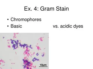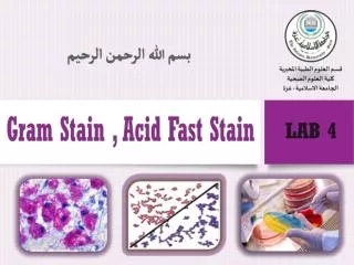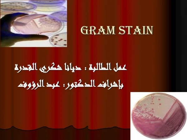
Gram Stain
Gram Stain. Gram Stain Reaction. The Gram stain reaction, named after the Danish bacteriologist Hans Christian Gram, was originally devised in 1882. Gram staining is based on the ability of the bacterial cell wall to retain the crystal violet dye.


Gram Stain
E N D
Presentation Transcript
Gram Stain Reaction • The Gram stain reaction, named after the Danish bacteriologist Hans Christian Gram, was originally devised in 1882. • Gram staining is based on the ability of the bacterial cell wall to retain the crystal violet dye. • Whether the crystal violet dye is retained or not is dependent on the type of bacterial cell.
Two Categories of Bacteria • Gram Positive • Gram Negative
Gram Negative vs. Gram Positive Bacteria Gram positive bacteria differ from Gram negative bacteria in the structure of their cell walls. The cell walls of Gram positive bacteria are made up of 20x more peptidoglycan, a polymer made of sugar and amino acids.
Gram Stain Process Gram Positive Bacteria • Step 1: Heat fix the slide so that that bacterial cells are fixed to the slide. • Step 2: Flood the slide with crystal violet dye. • The individual crystal violet ions penetrate the thick peptidoglycan layer of the cell as well as the plasma membrane. Gram Negative Bacteria • Step 1: Heat fix the slide so that that bacterial cells are fixed to the slide. • Step 2: Flood the slide with crystal violet dye. • The individual crystal violet ions penetrate the thin peptidoglycan layer of the cell as well as the plasma membrane.
Gram Stain Process Continued Gram Positive Bacteria • Step 3: Rinse with water. • Step 4: Flood the slide with iodine. • The iodine ions penetrate the cell wall and bind with the crystal violet. • The iodine ions and crystal violet react, forming a crystal violet-iodine complex that is a very large molecule and is insoluble in water. Gram Negative Bacteria • Step 3: Rinse with water. • Step 4: Flood the slide with iodine. • The iodine ions penetrate the cell wall and bind with the crystal violet. • The iodine ions and crystal violet react, forming a crystal violet-iodine complex that is a very large molecule and is insoluble in water.
Gram Stain Process Continued Gram Positive Bacteria • Step 5: Rinse with water. • Step 6: Decolorize the slide with alcohol. • The alcohol causes water to leave the cell wall. Because of its larger size, the crystal violet-iodine complex is blocked from moving easily through the cell wall and thus is prevented from leaving the cell. Gram Negative Bacteria • Step 5: Rinse with water. • Step 6: Decolorize the slide with alcohol. • The alcohol disrupts and dissolves the outer membrane. Therefore the big crystal violet-iodine complex is able to leave the cell.
Gram Stain Process Continued Gram Positive Bacteria • Step 7: Rinse with water. • The crystal violet-iodine complex is still stuck inside the cell. The cell is therefore stained purple. • Step 8: Flood the slide with Safranin dye. Gram Negative Bacteria • Step 7: Rinse with water. • The crystal violet-iodine complex is washed away, leaving colorless, unstained cells. • Step 8: Flood the slide with Safranindye.
Gram Stain Process Continued Gram Positive Bacteria • Safranin penetrates the cell, but since it is a lighter color than the crystal violet-iodine complex, it is not visualized over the purple color of the crystal violet-iodine. • The cells appear purple. Gram Negative Bacteria • Safranin penetrates the cell and stains the cell a reddish-pink color. • The cells appear reddish-pink.