Background
Jussi Tallus 1 , Pantelis Lioumis 2 , Heikki Hämäläinen 3 , Seppo Kähkönen 2 , Olli Tenovuo* 1 1 Department of Neurology, University of Turku, Finland, 2 Biomag Laboratory, Finland, 3 Department of Psychology, University of Turku, Finland *presenting author.

Background
E N D
Presentation Transcript
Jussi Tallus 1, Pantelis Lioumis2, Heikki Hämäläinen3, Seppo Kähkönen2, Olli Tenovuo*11Department of Neurology, University of Turku, Finland, 2Biomag Laboratory, Finland, 3Department of Psychology, University of Turku, Finland *presenting author TMS-evoked EEG responses in symptomatic and recoveredpatientswithmildtraumaticbraininjury
Background • Mostpatientsrecoverfrommildtraumaticbraininjury (mTBI) within 3 months • About 15% developpersistentsymptoms • Diffuseneuronaldamage and anteriorbraintractdysfunctionareprobablyinvolved in at leastsomemTBIcaseswithchronicsequels (according to diffusiontensorimaging and fMRIstudies)
TMS-EEG • Transcranialmagneticstimulation (TMS): A rapidlychangingmagneticfieldinduces a brief, focalelectricfield in the brain • TMS with EEG enablesreal-timemeasurement of brainresponses to standardizedstimulation • Excitationoccurs on corticalgraymatter, butcortico-cortical and thalamo-corticalconnectionsmodify the activity of the stimulatedcircuits
Participants • 19 mTBI (GCS 13-15) patients, 11 withpersistentsymptoms and 8 fullyrecovered • 9 healthycontrols • No signs of injury in MRI in visualinspection • No CNS affectingmedications
Experimentalprocedure • Single pulse TMS on leftdorsolateralprefrontalcortex (DLPFC) and leftprimarymotorcortex (M1) in trains of 100 pulses (at 0.3Hz) • Motor thresholds (MTs) measuredusing EMG, stimulus trainsappliedwithintensities 90%, 100%, and 110% MT in randomizedorder
Data analysis • Responsesaveraged for everystimulationcondition • Pools of 4-6 electrodescreated for 8 regions of interest: left and righthemisphereprefrontalcortex (LPFC & RPFC), motorcortex (LMC & RMC), temporalcortex (LTC & RTC), and parietalcortex (LPC & RPC)
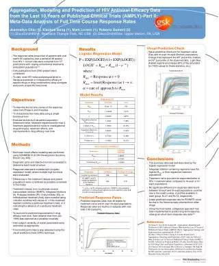
Results • In all groups, the same peaks were identified in the TMS–evoked EEG responses. • Statistically significant differences between the groups in peak amplitudes and latencies were observed in all time ranges, especially P30 and N100 deflections, and later in P200, P300, and the 370-440 ms time range mean amplitude.
Results • Differences in the earlier deflections were mostly seen in the frontal areas.
P30-N100 Recoveredvssymptomatic
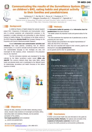
Results • Differences in the late (P300, 370-440 ms mean amplitude) time range were most pronounced in the temporal and parietal electrodes.
300-500 ms Controls vspatients
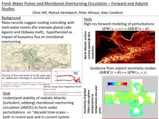
Results • Patients in the symptomatic group were found to miss one or more of the normally appearing peaks more often than controls (p = 0.024). Control SymptomaticmTBI SymptomaticmTBI
N100-P200 deflection Recoveredvssymptomatic
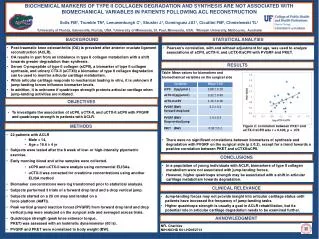
1 symptomatic 2 recovered 3 control Function 2 Group centroid 3 2 1 Function 1 Stepwiselineardiscriminantanalysis of allthreegroups: 85.7% correctlygrouped. Variablesincluded: DLPFC 90% MT rightparietal and rightprefrontal P30 amplitudes, M1 90% MT lefttemporal P300 amplitude, and DLPFC 110% MT rightprefrontal N100 amplitude.
Conclusions Based on currentknowledge, interpretation of the observedchangesseen is uncertain: • P30: mayberelated to an earlyspreading of activation to functionallyconnectedareas (longerlatencies in the symptomaticsubjects) • N100: thought to reflect a corticalinhibitoryprocesstriggeredby TMS (more negative in the symptomatic group on prefrontal areas) • P300 and later: currentknowledgeverylimited, highercognitiveprocesses? (consistentlylowest in the symptomaticsubjects)
Conclusions • A validated combination of TMS-evoked EEG responses could be valuable in separating mTBI subjects from controls and recovered mTBI subjects from those with chronic sequels (LDA of the two patient groups with three best variables grouped correctly 100% of cases)