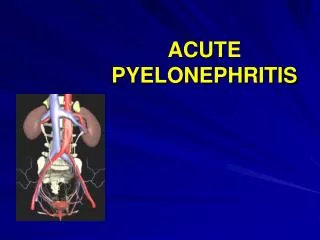
ACUTE PYELONEPHRITIS
ACUTE PYELONEPHRITIS. ACUTE PYELONEPHRITIS. The acute pyelonephritis is a nonspecific infectious disease that involves the pelvis of the kidney, calyces and parenchyma, particularly its interstitial tissue. 20-40% of the patients with the renal diseases suffers of the pyelonephritis.

ACUTE PYELONEPHRITIS
E N D
Presentation Transcript
ACUTE PYELONEPHRITIS • The acute pyelonephritis is a nonspecific infectious disease that involves the pelvis of the kidney, calyces and parenchyma, particularly its interstitial tissue. • 20-40% of the patients with the renal diseases suffers of the pyelonephritis.
ACUTE PYELONEPHRITIS • Approximately 250,000 cases of acute pyelonephritis occur each year, resulting in more than 100,000 hospitalizations. • Women are approximately five times more likely than men to be hospitalized with this condition (11.7 versus 2.4 hospitalizations per 10,000 cases, respectively); however, women have a lower mortality rate than men (7.3 versus 16.5 deaths per 1,000 cases, respectively)
Microbial Organisms Causing Specific Types of Urinary Tract Infections
Pathogenesis • Renal infection usually ascends from the urethra and lower genitourinary tract. Hematogenous infection of the kidney occurs infrequently; lymphatic spread occurs rarely, if ever. • The short urethra in girls and women and its close proximity to the anus allow periurethral pathogenic bacteria easy access to the bladder during sexual intercourse or urethral manipulation. • Once bladder infection is established, whether infection ascends via the ureters and involves the kidneys is influenced by microbial virulence factors, the presence or absence of vesicoureteral reflux, the quality of ureteral peristalsis, and the susceptibility of the renal medulla to infection.
Pathogenesis • A. Hematogenous spread. Infections spread from the distant place /otitis, tonsillitis, bronchitis, pneumonia, osteomyelitis, mastitis, furunculosis, wounds/. • B. Urinogenic spread. Infection goes at the ureter from the bladder because of the vesicoureteral reflux. The pelvicovenous, pelvicolymphathic, fornical and tubular refluxes may occur. • C. Ascending infection. Ascending infection from the ureter at the subepithelial tissue passes direct into the interstitial tissue of the kidney. • D. Lymphatogenous spread. The infection by means of the lymphatic channels probably occurs but it is rare. The promote factors are general /avitaminosis, supercooling, overheating, other infection diseases, gastric ulcer, etc./ and local /intravesical obstruction, neuromuscular dysplasia of ureter, benign prostate hyperplasia, etc/.
Pathogenesis • Urinogenic spread. Infection goes at the ureter from the bladder because of the vesicoureteral reflux.
Classification • The primary and secondary pyelonephritis are distinguished. • There is no dysfunction of the urine outflow during the primary pyelonephritis. • The secondary pyelonephritis goes with urostasis.
Classification • 1/ The unilateral and bilateral. • a/ Acute /purulent, serous/ • b/ Chronic; • c/ Relapsing course. • 2/ By the mode of bacteria pathway there are differed: • 3/ a/ hematogenous /ascending/; • b/ urogenic /ascending/; • c/ urolithiasis /infected urinary stones/; • d/ tuberculosis of the kidneys; • e/ the other renal diseases. • By the course, age, stage of the organism there are differed: • 1/ the pyelonephritis of newborn; • 2/ the pyelonephritis of the aged patients; • 3/ the pyelonephritis of the pregnant women; • 4/ the pyelonephritis in diabetes mellitus patients. • The acute pyelonephritis may be complicated with purulent nephritis, carbuncle of the kidney, the renal abscess, renal insufficiency.
Clinical Findings • A.Symptoms: • Symptoms of cystitis: frequency, nocturia, urgency, and dysuria. • Significant malaise and prostration are the rule; nausea, vomiting, and even diarrhea are common. • Young children most often complain of poorly localized abdominal discomfort and seldom localize the discomfort specifically to the flank.
Clinical Findings • B.Signs: • The patient generally appears quite ill. Intermittent chills are associated with fever ranging from 38.5 to 40 °C (101-104 °F) and tachycardia (the pulse rate may range from 90/min to 140/min or faster). • Fist percussion over the costovertebral angle overlying the affected kidney usually causes pain.
Laboratory Findings • The hemogram typically shows significant leukocytosis; the erythrocyte sedimentation rate is increased. • Urinalysis usually shows cloudy fluid with heavy pyuria, bacteriuria, mild proteinuria, and often microscopic or gross hematuria. • Urine cultures are positive in 90 percent of patients with acute pyelonephritis, and cultures should be obtained before antibiotics therapy is initiated. • The use of blood cultures should be reserved for patients with an uncertain diagnosis, those who are immunocompromised, and those who are suspected of having hematogenous infections
Ultrasonography • Ultrasonography shows flattering and dilatation of the calyces and renal pelvis, dysfunction of the urine passages, edema of the adipose capsule looks as rarefaction about the kidney. • It also shows the sizes of the concrement in the kidney.
Ultrasonography • Dilated pelvicaliceal system in a female patient with acute onset of flank pain radiating to the genitalia.
X-Ray Findings • A plain film of the abdomen may show some degree of obliteration of the renal outline owing to edema of the kidney and perinephric fat. • The outline of the ileopsoas muscle is absent sometimes, the diffuse shadow about the kidney and moderate scoliosis at the side of the disease are present. • Suspicious calcifications, must be carefully evaluated, because infected renal stones and calculous obstruction complicating pyelonephritis require special management.
X-Ray Findings • The excretory urograms show the enlargement of the infected kidney. • There is a slow excretion of the contrast. Calyces are flattered and clubbed, they are filled with contrast later than normal kidney. • The intravenous excretory urogram shows the significant atrophy of the parenchyma of the affected kidney, its deformation because of infiltrates and atonia of the ureter.
Instrumental Examination • There can be seen the bullous edema of the urethral orifice because of calculus at the intravesical portion, ureterocele, tumor compression. • Chromocystoscopia shows the range even sometimes the cause of the functional loss of the urine outflow.
Differential Diagnosis • The differential diagnosis of acute pyelonephritis includes pelvic inflammatory disease, cholecystitis, appendicitis, lower lobe pneumonia, perforated viscus, and the prodrome of herpes zoster.
Treatment • A.Specific Measures • B. General Measures • C.Failure of Response • D. Follow-Up Care
Treatment • Specific Measures: • When the infection is severe or complicating factors are present, hospitalization may be required. • Urine and blood specimens must be obtained immediately for culture; recognized pathogens must be tested for antimicrobial sensitivity. • Until the results of these tests are known, antimicrobial drugs should be given empirically.
Indications for Hospitalization in Patients with Acute Pyelonephritis
Treatment • General Measures: Complete bed rest is advised until symptoms subside. • Medication should be given for pain, fever, and nausea. • It is important to give fluids intravenously and orally to ensure adequate hydration and maintenance of adequate urinary output.
Treatment • Antimicrobial treatment with desintoxicative and general stimulate measures are effective in case of primary process. • An acute primary pyelonephritis is treated massively with the maximal dosage of the antibacterial mediums in different combinations. • The secondary pyelonephritis requires draining of the kidney, sometimes even the purulent source removal. Before the urine outflow isn’t restored the antibacterial mediums are dangerous especially of the strong action. The bacteriemic shock may develop.
Antimicrobial Agents Used in the Treatment of Acute Pyelonephritis
Acute purulent pyelonephritis • Apostematous pyelonephritis • Renal carbuncle • Abscesses of the kidney
Apostematous pyelonephritis • It is the suppuration of the parenchyma of the kidney with formation of the small multiply purulent focuses /apostems/. • The process occurs unilateral and bilateral. The purulent focuses are direct under the fibrous capsule, 1-3 mm in size and merging sometimes.
Renal abscess • Renal cortical abscesses develop primarily as a result of hematogenous spread of Staphylococcus aureus infections at distant sites, most often the skin (furunculosis, felon, mastitis). • At times, foci of primary renal infections caused mainly by gram-negative bacteria (coliform organisms) coalesce in the renal medulla to form abscesses. • In the past, most renal abscesses were caused by staphylococci; recently, coliform bacteria have become the predominant pathogens in renal abscesses.
Renal carbuncle • It is the suppurate-necrotic damage with formation of the bordered infiltrate in the cortex of the kidney. • Renal cortical carbuncles develop primary as a result of the hematogenous spread of infection form the distant sites /most often from the respiratory tract, suppurative diseases of kidney, skin, furunculosis, felon, mastitis, etc./ • Mechanism of carbuncle formation is the septic embolism of the renal artery that causes the septic infarction of the kidney and the development of carbuncle.
Clinical Findings • A.Symptoms: renal abscess is typified by an abrupt onset of chills, fever, and localized costovertebral pain. In the early stages, when the abscess does not communicate with the collecting system, symptoms of vesical irritability are absent and urinalysis is normal, although the patient may appear quite septic. The clinical picture often mimics that of acute pyelonephritis. • B.Signs: In acute cases, localizing signs are flank tenderness, possibly a palpable mass, and erythema and edema of the skin of the overlying loin. At times, however, abscesses associated with both acute and chronic infections present as febrile illnesses with few localizing signs.
Laboratory Findings • The hemogram usually shows marked leukocytosis with a shift to the left. • With cortical abscesses that do not communicate with the collecting system, urinalysis shows no pyuria or bacteriuria, and urine culture is negative. • Medullary abscesses generally are associated with heavy pyuria, bacteriuria, and positive urine cultures. • Blood cultures may be positive. • Depending upon the extent of renal involvement and associated renal abnormalities, the serum creatinine and urea nitrogen values may be normal or elevated.
X-Ray Findings • If the renal outline is visible, the plain film may show an enlarged kidney or a bulge of the external renal contour. With perinephric edema, however, often the renal outline is obliterated and the psoas shadow indistinct. • Unless the abscess has ruptured into the perinephric space or is quite large, scoliosis generally is not observed. Renal stones may be noted.
X-Ray Findings • When cortical abscesses are small, the excretory urogram may appear normal; most often, however, a space-occupying lesion (the abscess) is delineated. • Pyelonephritic changes, hydronephrosis, and urolithiasis also may be observed.
X-Ray Findings • Pyelonephritic changes, hydronephrosis, and urolithiasis also may be observed.
Ultrasonography • Intrarenal abscesses appear as well-marginated round or oval masses with irregular walls on ultrasonography. • The mass may be echo-free or slightly echogenic due to the presence of necrotic debris.
Treatment /Apostematous Pyelonephritis/ • The urgent surgical measures are required. • The subcostal lumbotomia is performed. The kidney is nude and decapsulated. The purulent focuses are incised. The retroperitoneal space should be drained and free output of the urine provided by means of the nephrostomy. • The surgical drainage should be present until the urine output becomes free, inflammation disappears and renal function normalizes. • The postoperative period requires the antibacterial and desintoxicative treatment that is similar to chronic pyelonephritis.
Treatment • Renal carbuncle • Lumbotomia is performed. Then decapsulation of the kidney and cone-shaped excision of the carbuncle are done. • Incision, curettage and draining of the kidney or enucleating of the carbuncle with its incision may be performed too. • Nephrectomy and partial nephrectomy are required less often today than in the past.
Gestation pyelonephritis • The inflammatory process develops while pregnancy, delivery and puerperal period.
Gestation pyelonephritis(Pyelonephritis of pregnancy) • Most frequently it is observed in pregnant (48%) more rare in puerperal (35%) women. • It develops while 1 pregnancy 2 trimester often. • There are women 18-25 years old.
Etiology /Pyelonephritis of pregnancy/ • Urinoculture finds out E.Coli, Staphylococcus albicans, Clebsiella in pregnant women. • Association of the Proteus and Blue pus bacilli is observed in puerperal women. • The primary source of the infection may be any purulent inflammatory place (furunculosis, dental caries, inflammatory diseases of the genital organs).
Pathogenesis/Pyelonephritis of pregnancy/ • The pathogenetic sign is bacteriuria. It is observed in 7% only. 20-40% of women with asymptomatic bacteriuria develop pyelonephritis during pregnancy. Treatment of the bacteriuria lowers this risk. • Urodynamic dysfunction favors the pyelonephritis development. • Pathogenesis may be explained with mechanical, neurohumoral and endocrine factors. • The enlarged uterus compresses the pelvic portion of the ureters causing ureteropyeloectasia while pregnancy. • The moderate hypotonia and hypokinesia of the calicopelvic of the both kidneys and ureters are observed on 8th week. • Changes of the upper portion of the urinary tract may be explained by weakening of the sympathetic nervous system tonus. • Dysfunction of the urinary output because of the urinary pathway atonia is a condition for pathogen activation. Vesicoureteral and pelvicorenal refluxes favor spreading of the infection into the interstitial tissue of the renal parenchyma.
Clinical findings /Pyelonephritis of pregnancy/ • Clinical findings have the own peculiarities according to the different terms of pregnancy. They also depend on the range of the urinary output damage. • A sharp pain in loin that irradiates to the lower portions of the abdomen, genitals are at the 1 trimester. • 2nd and 3rd trimesters are characterized with a moderate pain because of the dilatation of the upper urinary tract and intrarenal pressure decreasing. • An acute purulent pyelonephritis develops more frequently in pregnant and postpueral women.
Clinical findings /Pyelonephritis of pregnancy/ • Symptoms: • The usual symptoms of acute pregnancy pyelonephritis include abrupt onset of shaking chills, moderate to high fever, a constant ache in the loin (unilateral or bilateral). • Symptoms of cystitis: frequency, urgency, and dysuria. • Significant malaise and prostration are the rule; nausea, vomiting.
Diagnosis/Pyelonephritis of pregnancy/ • The enlarged uterine hinders the palpation • Laboratory Findings • Chromocystoscopia • Ultrasonography