Download
1 / 61
650 likes | 824 Vues
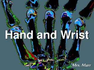
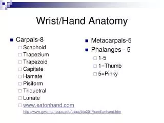
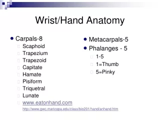
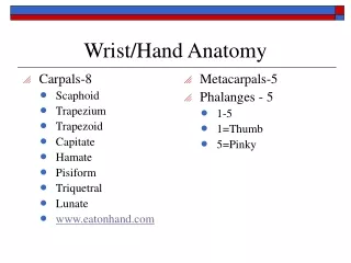
Anatomy of the Hand and Wrist. Mnemonic for Learning Carpals. S he L ikes T o P lay. Lunate In the moonlight. Scaphoid A boat. Triquetrum The third T Bone. Pisiform Pea-shaped. Hamate A hambone With a hook. Trapezium: “It’s by the thumb”. Capitate. Trapezoid “Is by its side”.

E N D
MnemonicforLearningCarpals She Likes To Play Lunate In the moonlight Scaphoid A boat Triquetrum The third T Bone Pisiform Pea-shaped Hamate A hambone With a hook Trapezium: “It’s by the thumb” Capitate Trapezoid “Is by its side” Try To Catch Her Click R Button for Slideshow
Flexor Tendons • The muscles that flex your wrist are on the palmer side. • A group of the begin at the medial epicondyle of the humerus at the elbow
Flexor Carpi Radialis • Origin Medial epicondyle of humerus • Insertion Base of 2nd metacarpal • Action Flexes and radial deviates the hand (at wrist) • Innervation Median nerve (C6 and C7)
Flexor Digitorum Superficialis • Origin medial epicondyle of humerus, • Insertion middle phalanges of digits 2 - 5 • Action Flexes middle phalanges at proximal interphalangeal joints also flexes proximal phalanges at metacarpophalangeal joints and hand • Innervation Median nerve (C7, C8 and T1)
Flexor Carpi Ulnaris • Origin medial epicondyle of humerus • Insertion Pisiform bone, hook of hamate bone, and 5th metacarpal bone • Action Flexes and ulnar deviates hand (at wrist) • Innervation Ulnar nerve (C7 and C8)
Palmaris Longus • Origin Medial epicondyle of humerus • Insertion Distal half of flexor retinaculum and palmar aponeurosis • Action Flexes hand at the wrist • Innervation Median nerve (C7 and C8)
Flexor Digitorum Profundus • Origin Proximal 3/4 of ulna • Insertion Base of the distal phalanx of digits 2 – 5 • Action Flexes distal phalanges at distal interphalangeal joints • Innervation • Medial part: ulnar nerveLateral part: median nerve
Flexor Digiti Minimi Brevis • Origin Hook of hamate and flexor retinaculum • Insertion Medial side of base of proximal phalanx of little finger • Action Flexes proximal phalanx of little (5th) finger • Innervation ulnar nerve
Flexor Pollicis Brevis • Origin Flexor retinaculum and tubercles of scaphoid and trapezium • Insertion Lateral side of base of proximal phalanx of thumb • Action Flexes thumb • Innervation Recurrent branch of median nerve (C8 and T1)
Flexor Pollicis Longus • Origin Anterior surface of radius and adjacent interosseous membrane • Insertion Base of distal phalanx of thumb • Action Flexes phalanges of 1st digit (thumb) • Innervation Anterior interosseous nerve from median nerve (C8 and T1)
The 2 Muscles of Pronation • Two muscles work together to turn the radius over the ulna and put the hand in a prone position • Pronator teres and pronator quadratus
Pronator Quadratus • Origin Distal 1/4 of anterior surface of ulna • Insertion Distal 1/4 of anterior surface of radius • Action Pronates forearm; • Innervation median nerve
Pronator Teres • Origin Medial epicondyle of humerus • Insertion Middle of lateral surface of radius • Action Pronates • Innervation Median nerve (C6 and C7)
Abduction at Hand • Abduction really only occurs at the thumb and little fingers
Abductor Digiti Minimi • Origin Pisiform • Insertion Medial side of base of proximal phalanx of little finger • Action Abducts little (5th) finger • Innervation ulnar nerve (C8 and T1)
Abductor Pollicis Brevis • Origin scaphoid and trapezium • Insertion Lateral side of base of proximal phalanx of thumb • Action Abducts thumb • Innervation median nerve (C8 and T1)
Abductor Pollicis Longus • Origin Posterior surfaces of ulna, • Insertion Base of 1st metacarpal • Action Abducts thumb • Innervation the radial nerve
Adduction • Movement towards the midline of the body
Adductor Pollicis • Origin 2nd and 3rd metacarpals, capitate, • Insertion Medial side of base of proximal phalanx of thumb • Action Adducts thumb • Innervation ulnar nerve
Wrist Extensors • The extensors of the wrist are on the Dorsal side of the forearm • A majority of the wrist extensors begin at the lateral epicondyle
Extensor Carpi Radialis Brevis • Origin Lateral epicondyle of humerus • Insertion Base of 3rd metacarpal • Action Extends and radially deviates the wrist • Innervation radial nerve (C7 and C8)
Extensor Carpi Radialis Longus • Origin Lateral supracondyle ridge of humerus • Insertion Base of 2nd metacarpal • Action Extends and radially deviates at the wrist • Innervation Radial nerve (C6 and C7)
Extensor Carpi Ulnaris • Origin Lateral epicondyle of humerus • Insertion Base of 5th metacarpal • Action Extends and ulnar deviates hand at wrist joint • Innervation Radial nerve
Extensor Digiti Minimi Origin Lateral epicondyle of humerus Insertion 5th digit Action Extends 5th digit at metacarpophalangeal and interphalangeal joints Innervation Posterior interosseous nerve
Extensor Digitorum • Origin Lateral epicondyle of humerus • Insertion Extensor expansions of medial four digits • Action Extends the four digits and the wrist • Innervation Posterior interosseous nerve
Extensor Indicis • Origin Posterior sufrace of ulna and interosseous membrane • Insertion Extensor expansion of 2nd digit • Action Extends 2nd digit and helps to extend hand • Innervation Posterior interosseous nerve
Extensor Pollicis Brevis • Origin Posterior sufraces of radius and interosseous membrane • Insertion Base of proximal phalanx of thumb • Action Extends proximal phalanx of thumb at carpometacarpal joint • Innervation Posterior interosseous nerve
Extensor Pollicis Longus • Origin Posterior surface of middle 1/3 of ulna • Insertion Base of distal phalanx of thumb • Action Extends distal phalanx of thumb at carpometacarpal and interphalangeal joints • Innervation Posterior interosseous nerve
Supination Muscles • There are two muscles that return you to the anatomical position by uncrossing the radius and ulna
Biceps Brachii • Origin • Short head: tip of coracoid process of scapulaLong head: supraglenoid tubercle of scapula • Insertion Tuberosity of radius and fascia of forearm via bicipital aponeurosis • Action Supinates forearm and, when it is supine, flexes forearm • Innervation Musculocutaneous nerve (C5 and C6 )
Supinator • Origin Lateral epicondyle of humerus, • Insertion Lateral, posterior and anterior surfaces of proximal 1/3 of radius • Action Supinates forearm • Innervation Deep branch of radial nerve (C5 and C6)
Opposition • The ability to touch your thumb and pinky
Opponens Digiti Minimi • Origin Hook of hamate and flexor retinaculum • Insertion Medial border of 5th metacarpal • Action brings little finger (5th digit) into opposition with thumb • Innervation Deep branch of ulnar nerve (C8 and T1)
Opponens Pollicis • Origin Flexor retinaculum and tubercles of scaphoid and trapezium • Insertion Lateral side of 1st metacarpal • Action Draws 1st metacarpal laterally to oppose thumb toward center of palm • Innervation Recurrent branch of median nerve (C8 and T1)
The median nerve • The median nerve supplies feeling the the palmer side of your 1st, 2nd,3rd, and medial 4th fingers. • The Median nerve is involved with carpal tunnel syndrome
The Ulnar nerve • The Ulnar nerve supplies feeling and motor function to the lateral 4th and 5th fingers.
The Radial Nerve • The radial nerve innervates most of the extensors and supplies the feeling on the dorsal side of the first three digits
Ball and socket joint – greatest range of motion allowing bones to swing in a circle Example: shoulder or hip
Pivot joint – one bone rotates around another Example: Neck and under the Elbow
Hinge joint – bones bend like a hinge forward and backward Example: Knee and Elbow
Gliding joint – allows one bone to slide over another Example: Wrist and Ankle
Joint Shapes • Condyloid: egg-shape articular surface + oval concavity • side-to-side, back+forth movement • (eg) metacarpophalangeal (knuckle)
pg 225 Joint Shapes • Saddle: articular surface both concave + convex • side-to-side, back-forth movement • (eg) carpometacarpal jt of thumb
Hinge Joint • In between the Phalanges are Hinge Joints • They move in flexion and extension
A Ligament • A Ligament attaches a bone to bone • An Injury to a Ligament is called a Sprain • A Tendon Attaches a muscle to a bone • An injury to a tendon or Muscle is a strain