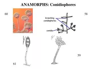
ANAMORPHS: Conidiophores
ANAMORPHS: Conidiophores. 60. 58. 59. 61. ANAMORPHS: Hyphomycetes and Coelomycetes. 63. 62. 63. 63. ANAMORPHS: Blastic versus Thallic Conidiogenesis. Fig. 8.6 in Webster and Weaver 2007. ANAMORPH: Holoblastic versus Enteroblastic Conidiogenesis. Fig. 8.6 in Webster and Weber 2007.

ANAMORPHS: Conidiophores
E N D
Presentation Transcript
ANAMORPHS: Conidiophores 60 58 59 61
ANAMORPHS: Hyphomycetes and Coelomycetes 63 62 63 63
ANAMORPHS: Blastic versus Thallic Conidiogenesis Fig. 8.6 in Webster and Weaver 2007
ANAMORPH: Holoblastic versus Enteroblastic Conidiogenesis Fig. 8.6 in Webster and Weber 2007
ANAMORPHS: Phialidic Enteroblastic and Annelidic Enteroblastic Conidiogenesis Fig. 8.7 in Webster and Weaver 2007
ANAMORPHS: Holothallic vs. Arthric-Thallic Conidiogenesis Fig. 8.0 in Webster and Weber 2007
ANAMORPHS: Schizolytic vs. Rhexolytic Secession (= Dehiscence) Fig. 4.5 in Kendrick 2000
ANAMORPHS: Conidiospore Shapes Fig. 8-2 in Alexopoulos et al. 1996
Powdery Mildew Oidium
122 Oidium 124
Histoplasmosis (“Spelunker’s Disease”) 72 Histoplasma 71
67 70 Histoplasma 68
Athlete’s Foot; Ringworm; Jock Itch 75 78 74 76 73 Trichophyton
107 106 Nematode-Trapping Fungus (Arthrobotrys)
Sticky Knobs Arthrobotrys Fig. 25.1 in Webster and Weber 2007
Adhesive Nets Arthrobotrys Fig. 25.2 in Webster and Weber
Constricting Rings Arthrobotrys Fig. 25.6 in Webster and Weber 2007
82 81 Penicillium (Eupenicillium)
92 Penicillium
86 87 88 89 Penicillium
Alexander Fleming 83 85 84 Penicillium
93 94 Aspergillus (Eurotium)
96 97 Koji Sake 89 110
95 111 Aspergillosis 112
Dutch Elm Disease 100 101
Dutch Elm Disease Ophiostoma ulmi 99 98
Pine Bark Beetle Damage 104 Blue Stain Fungus (Ophiostoma minus) 103 102
Canker, Coral Spot Nectria
Sporodochia Nectria
Anthracnose Colletotrichum
Acervuli 115 116 117 114 Colletotrichum
1 http://www.caf.wvu.edu/kearneysville/disease_descriptions/ disease_images/phot2-55.jpg 2 http://botit.botany.wisc.edu/images/332/Ascomycota/H emiascomycetes/Taphrina_deformans_asci_tjv.low.jpg 3 http://www.mycolog.com/4_Taphrina_deformans2.jpg 4 http://www.naturephoto-cz.com/photos/maly/peziza-badia-174.jpg 5 http://ocid.nacse.org/classroom/fungi/bot461/images/peziza2.jpg 6 http://www.eou.edu/~kantell/img1015.jpg 7 http://mycoweb.narod.ru/fungi/Submitted/ESP/Ascobolus_furfuraceus_ESP.jpg 8 www.mycolog.com/11-16_Ascobolus3.jpg 9 http://www.uoguelph.ca/~gbarron/MISCELLANEOUS/ascobolu.htm 10 http://www.villaohiggins.com/blog/uploaded_images/Morchella_esculenta -722783.jpg
11 http://www.hviidphotography.dk/b/uploaded_images/ morchella-735723.jpg 12 http://www.ereleases.com/pr/2006-MorelFarms.jpg 13 http://morelmushroomhunting.com/morelfindsssss.jpg 14 http://www.ereleases.com/pr/2006-MorelFarms.jpg 15 http://www.fmnh.helsinki.fi/users/harmaja/Gyromitra.jpg 16 http://weblogs.variety.com/photos/uncategorized/wine_tours_truffle_1.jpg 17 http://bugs.bio.usyd.edu.au/Mycology/images/Topics/Reprodn_ Dispersal/truffleTuber.jpg 18 http://www.markys.com/caviar/customer/image.php?type=P&id=18036 20 http://www.fao.org/docrep/007/Y5489E/y5489e-12.jpg 21 http://www.fao.org/docrep/007/Y5489E/y5489e-10.jpg 22 https://www.whitetruffleauction.com/shop/images/Prod_BlackWin30g.jpg
23 http://farm4.static.flickr.com/3165/2893140294_5759b8855b.jpg?v=0 24http://botit.botany.wisc.edu/toms_fungi/images/xypericl.jpg 25 http://www.fungi4schools.org/Reprints/Photoset01/Xylaria_hypoxylon_ candle_snuff_fungus-01.jpg 26 http://www.north-west-wildlife.co.uk/Fungi%20Photo3.htm 27 http://www.messiah.edu/Oakes/fungi_on_wood/club%20and%20coral/ images/Xylaria%20hypoxylon%20close%20up%20DW.jpg 28 http://www.messiah.edu/Oakes/fungi_on_wood/club%20and%20coral/i mages/Xylaria%20hypoxylon%20close%20up%20DW.jpg 29 http://www.botanik.uni-karlsruhe.de/garten/fotos-knoch/Claviceps%20purpurea%20Mutterkorn%201.jpg 30 http://www.erowid.org/plants/ergot/images/archive/claviceps_purpurea3.jpg 31 http://www.myko.cz/include/slovnik/images/ana-teleomorfa.gif
31 http://www2.biomed.cas.cz/~pazouto/images/citrina.gif 32 http://www.dipbot.unict.it/sistematica/Immagini/13001.JPG 33 http://www.myko.cz/include/slovnik/images/ana-teleomorfa.gif 34 http://botit.botany.wisc.edu/images/332/Ascomycota/Plectomycetes/Asperg._ Eurotium_per._st._tjv.jpg 35 http://www.yates.com.au/images/au/problem-solver/peach-leaf-curl/peach-leaf-curl-1.jpg 36 http://media-2.web.britannica.com/eb-media/29/8729-004-CC77055F.jpg 37 http: ://botit.botany.wisc.edu/images/130/Fungi/Ascomycota_Images/ Peziza/Scarlet_cup.html&usg=__oyrd9unDXgKGB529BUAyoDVCFo=&h=358&w=480&sz=15&hl=en&start=1&tbnid=HVE3OUU-Jd1LiM:&tbnh=96&tbnw=129&prev=/ images%3Fq%3Dscarlet%2Bcup%26gbv%3D2%26hl%3Den%26sa%3DG 38 http://botit.botany.wisc.edu/toms_fungi/images/scab.jpg 39 http://www.caf.wvu.edu/kearneysville/disease_descriptions/disease_ images/phot2-45.jpg
40 http://botit.botany.wisc.edu/toms_fungi/images/scab.jpg 41 http://www.apsnet.org/education/LessonsPlantPath/AppleScab/images/ fig08.jpg 42 http:://www.checkout.org.cn/news/news_images.jsp%3Fcntn_id%3D 104205%26org%3DNSF&usg=__VrrPkjiip2ZXYBhJEHX2SIWzy7E=&h=743&w=687&sz=136&hl=en&start=3&tbnid=VftOAuUjukRFnM:&tbnh=141&tbnw=130&prev=/images%3Fq%3Dauxin%26gbv%3D2%26hl%3Den%26sa%3DG 43 http://www.uoguelph.ca/~gbarron/MISC2006/jul2001.jpg 44 http://www.istockphoto.com/file_thumbview_approve/470979/2/i stockphoto_470979-mouldy-stained-paper.jpg 45 http://www.csupomona.edu/~jcclark/classes/bot125/resource/&usg =__i6zPY-vrmPntteqtfXbuZj3QcjM=&h=178&w=150&sz=18&hl =en&start=49&tbnid=QihjatlfWLroM:&tbnh=101&tbnw=85&prev=/images%3Fq%3Dsordaria%26start%3D40%26gbv%3D2%26ndsp%3D20%26hl%3Den%26sa%3DN
46 http://www.stanford.edu/group/neurospora/RajuNcrassaWeb/Fig48.WTxcys3. Raju781216.gif 47 http://wards.scientificspot.com/wp-content/uploads/2007/07/figure1.gif 48 http://www.fgsc.net/2000compendium/figure1.jpg 49 http://www.fgsc.net/2000compendium/figure1.jpg 50 http://www.shroomery.org/images/23418/Lipstickmold.jpg 51 http://www.stanford.edu/group/neurospora/PhotoSeriesIntroGIF/ Fig9.Perithecia.Springer.gif 52 http://www.tolweb.org/tree/ToLimages/fig7tubeufiaascostroma.250a.jpg 53 http://www.fgsc.net/Neurospora/raju2_files/image004.jpg 54 http://staff.jccc.net/PDECELL/transgenetics/beasdtatum.gif 56 http://1.bp.blogspot.com/_DZH2cmCoois/SCsSgYuCcyI/AAAAAAAAFL4/ yaIcTx72uRw/s400/Nobel_Laureates_1958_Beadle_Tatum.bmp 57 http://www.palaeos.com/Fungi/Lists/Glossary/Images/Apothecium.jpg
58 http://www.fungionline.org.uk/6asexual/9conidioph.html&usg= __LAXrmmsPoA3YlFW3RAnfO_nwuoM=&h=144&w=504&sz=5&hl=en&start=12&tbnid=IE6jmHwiTB-y6M:&tbnh=37&tbnw=130&prev=/images%3Fq%3Dpycnidium%26gbv%3D2%26hl%3Den%26sa%3DG 59 http://www.scielo.br/img/revistas/abb/v19n3/27366f1.jpg 60 http://www.uoguelph.ca/~gbarron/MISCE2002/aspwen3.jpg 62 http://bugs.bio.usyd.edu.au/Mycology/images/Topics/Growth_Dev/synnemata.gif 61 http://ejournal.sinica.edu.tw/bbas/content/2000/3/1312030.JPG 64 http://www.fungionline.org.uk/images/6asexual/acer.gif 65 http://www.fungionline.org.uk/images/6asexual/pycnid.gif 66 http://www.mycolog.com/conidiogenesis%2012b.JPG 67 http://botit.botany.wisc.edu/toms_fungi/images/hcap1.jpg 68 http://www.doctorfungus.org/Mycoses/images/histoplasma1.jpg
69 http://www.pathguy.com/~tdemark/0073.jpg 70 http://www.sflorg.com/sciencenews/images/imscn042706_02_01.jpg 71 http://graphics8.nytimes.com/images/2007/08/01/health/adam/17222.jpg 72 http://depts.washington.edu/hivaids/images/oit/oit_c8_q01.jpg 73 http://www.provlab.ab.ca/mycol/image/derm/tpedis16.jpg 74 www.provlab.ab.ca/mycol/image/derm/toetrub.jpg 75 http://www.telmeds.org/AVIM/Amico/index_Amico_files/dermatofitos.jpg 76 http://pro.corbis.com/images/42-19880471.jpg?size=67&uid=%7BC9B37CEF- 65D3-4742-90F8-262588FF7F7A%7D 77 http://www.research.usf.edu/cm/pics/ringworm_1.jpg 78 http://www.allstop.com/images/examples/jock-itch4.gif 79 http://www.medmicro.wisc.edu/resources/imagelib/mycology/images/ trichophyton_rubrum.gif
80 http://library.thinkquest.org/C005271F/Penicillium3.jpg 81 http://img.photobucket.com/albums/v697/slaw/penicillium-roqueforti.jpg 82 http://www.bsu.edu/classes/ruch/msa/geiser/10.jpg 83 http://www.xtec.es/~jllort1/biolegseuropa/fleming.jpg 84 http://1.bp.blogspot.com/_1CRpQGsPBBg/RiMaLOvraNI/ AAAAAAAAAXg/fKWbFy4mKoo/s400/s5.gif 85 http://www.cartoonstock.com/lowres/cco0011l.jpg 86 http://www.thenibble.com/REVIEWS/MAIN/cheese/cheese2/images/ x-blue-230_000.jpg 87 http://pro.corbis.com/images/42-19880058.jpg?size=67&uid= %7B5024CC6C-193F-4C32-BB9C-5E374ECBA455%7D 88 http://www.skinsideout.co.uk/images/lipasebreakdown.jpg 89 http://www.servicexs.com/plaatjes/protease_reaction.jpg 90 http://webs.wichita.edu/mschneegurt/biol103/lecture23/rotting_spams.jpg
93 http://129.215.156.68/Images/Asexual%20structures%20of%20A spergillus%20niger.jpg 94 http://www.botany.hawaii.edu/faculty/wong/Bot201/Ascomycota/Asperg2.jpg 95 http://services.epnet.com/GetImage.aspx/getImage.aspx?ImageIID=7633 96 http://www.epicurious.com/images/articlesguides/drinking/wine/sakeMain3.jpg 97 http://www.tibbs-vision.com/sake/komegrainLres.jpg 98 http://www.apsnet.org/education/LessonsPlantPath/DutchElm/Images/fig07x.jpg 99 http://www.resista-ulmen.com/img/triple1.jpg 100 http://www.manitobaliberals.ca/uploaded_images/Iu19-763533.jpg 101 http://www.majestictreecare.com/Majestic_main/images/Photos/ElmLeafminer.jpg 102 http://everest.ento.vt.edu/~salom/SPBbiology/bluestain.html 103 http://www.genomealberta.ca/files/Images/blogs/Mountain_Pine_Beetle/G._ clavigera_conidiophore_-_web_small.jpg
104 http://www.ipm.ucdavis.edu/NEWS/IMAGES/barkbeetle.jpg 105 http://skyblu.files.wordpress.com/2006/11/mt-pine-bark-beetle.JPG 106 http://www.apsnet.org/education/LessonsPlantPath/LesionNema/Images/ fig18.jpg 107 http://www.biological-research.com/philip-jacobs%20BRIC/ 108 http://plpnemweb.ucdavis.edu/nemaplex/images/antago22.jpg 109 www.science-projects.com/starch.GIF 110 http://www.micro.siu.edu/micr201/images/Ethanol.gif 111 http://www.ispub.com/xml/journals/ijid/vol6n1/aspergillosis-fig9.jpg 112 http://pathhsw5m54.ucsf.edu/case16/images16/aspergilloma2.jpg 113 http://www.uoguelph.ca/~gbarron/MISC2003/arthrobo.htm&usg= __Rh2bcty9ClB0kb0FOQQpIHLiayU=&h=257&w=335&sz=12&hl=en&start=4 &tbnid=765UgO7vgJ3UXM:&tbnh=91&tbnw=119&prev=/images%3Fq%3D arthrobotrys%26hl%3Den%26sa%3DG
114 http://www.plantpath.wisc.edu/tddl/tddl/disimg/anth/acervuli.jpg 115 http://www.uoguelph.ca/~thsiang/turf/survey99/acervuli.jpg 116 http://ag.arizona.edu/plp/plpext/diseases/turf/warmseason/csanth3.jpg 117 http://www.bitterrootrestoration.com/images/botany/Diseases/ colletotrichum_gloeosporioides.jpg 118 http://www.ntbg.org/breadfruit/breadfruit/images/pests1.jpg 119 http://www.galeon.com/mundoagricola/fefa/Phoma/phoma1.jpg 120 http://mycoweb.narod.ru/fungi/Submitted/SJG4/Nectria_coccinea_2_JS_ 20070422.jpg 121 http://hcs.osu.edu/albums/SK_Notes/oidium.jpg 122 http://www.niaes.affrc.go.jp/inventory/microorg/eng/Oidium.jpg 123 http://www.bspp.org.uk/publications/molecular-plant-pathology/ pathprofiles/images/pathprofile12b.jpg