Download
1 / 30
440 likes | 982 Vues
IB Sports, exercise and health science. Movement analysis. Topic 4 Movement analysis. 4.1.1 Draw and label a diagram of a motor unit. Sub-topics. 1. Neuromuscular function. 2. Joint and movement type 3. Fundamentals of biomechanics.

E N D
IB Sports, exercise and health science Movement analysis Topic 4 Movement analysis 4.1.1 Draw and label a diagram of a motor unit. Sub-topics 1. Neuromuscular function 2. Joint and movement type 3. Fundamentals of biomechanics http://academic.wsc.edu/faculty/jatodd1/351/motor_unit.jpg
IB Sports, exercise and health science Movement analysis Topic 4 Movement analysis 4.1.1 Draw and label a diagram of a motor unit. Sub-topics 1. Neuromuscular function 2. Joint and movement type 3. Fundamentals of biomechanics
IB Sports, exercise and health science Movement analysis Topic 4 Movement analysis 4.1.1 Draw and label a diagram of a motor unit. Sub-topics 1. Neuromuscular function 2. Joint and movement type 3. Fundamentals of biomechanics
IB Sports, exercise and health science Movement analysis Topic 4 Movement analysis 4.1.1 Draw and label a diagram of a motor unit. Sub-topics 1. Neuromuscular function 2. Joint and movement type 3. Fundamentals of biomechanics http://www.brainexplorer.org/brain-images/dendrite.jpg
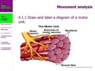
IB Sports, exercise and health science Movement analysis Topic 4 Movement analysis 4.1.1 Draw and label a diagram of a motor unit. Sub-topics • Label: dendrite, cell body, nucleus, axon, motor end plate, synapse and muscle. 1. Neuromuscular function 2. Joint and movement type 3. Fundamentals of biomechanics
IB Sports, exercise and health science Movement analysis Topic 4 Movement analysis 4.1.2 Explain the role of neurotransmitters in stimulating muscle contraction. Sub-topics • Neurotransmitters are chemicals that are used for communication between a neuron at the synapse and another cell. Wilmore et.al 2008 1. Neuromuscular function 2. Joint and movement type 3. Fundamentals of biomechanics
IB Sports, exercise and health science Movement analysis Topic 4 Movement analysis 4.1.2 Explain the role of neurotransmitters in stimulating muscle contraction. Sub-topics • Acetylcholine is the primary neurotransmitter for the motor neurons that innervate skeletal muscle and for most parasympathetic neurons. It is generally an excitatory neurotransmitter, but it can have inhibitory effects at some parasympathetic nerve endings, such as the heart. Wilmore et.al 2008 1. Neuromuscular function 2. Joint and movement type 3. Fundamentals of biomechanics
IB Sports, exercise and health science Movement analysis Topic 4 Movement analysis 4.1.2 Explain the role of neurotransmitters in stimulating muscle contraction. Sub-topics In biochemistry, cholinesterase is an enzyme that catalyzes the hydrolysis of the neurotransmitter acetylcholine into choline and acetic acid, a reaction necessary to allow a neuron to return to its resting state after activation. http://en.wikipedia.org/wiki/Cholinesterase_enzyme 1. Neuromuscular function 2. Joint and movement type 3. Fundamentals of biomechanics
IB Sports, exercise and health science Movement analysis Topic 4 Movement analysis 4.1.3 Explain how skeletal muscle contracts by the sliding filament theory. • When muscle contracts, muscle fibres shorten. How do they shorten? The explanation for this phenomenon is termed the Sliding Filament Theory. • When the myosin cross-bridges are activated, they bind with actin, resulting in a conformational change in the cross-bridge, which causes the myosin to tilt and to drag the thin filament toward the centre of the sarcomere. Wilmore et.al 2008 Sub-topics 1. Neuromuscular function 2. Joint and movement type 3. Fundamentals of biomechanics
IB Sports, exercise and health science Movement analysis Topic 4 Movement analysis 4.1.3 Explain how skeletal muscle contracts by the sliding filament theory. • This tilting of the head is referred to as the power stroke. The pulling of the thin filament past the thick filament shortens the sarcomere and generates force. When the fibres are not contracting, the myosin head remains in contact with the actin molecule, but the molecular bonding at the site is weakened or blocked by tropomyosin. (Tropomyosin is an actin-binding protein that regulates actin mechanics.) • Wilmore et.al 2008 Sub-topics 1. Neuromuscular function 2. Joint and movement type 3. Fundamentals of biomechanics
IB Sports, exercise and health science Movement analysis Topic 4 Movement analysis 4.1.3 Explain how skeletal muscle contracts by the sliding filament theory. • Troponin is a complex of three proteins that is integral to muscle contraction in skeletal and cardiac muscle, but not smooth muscle. Troponin is attached to the protein tropomyosin and lies within the groove between actin filaments in muscle tissue. In a relaxed muscle, tropomyosin blocks the attachment site for the myosin crossbridge, thus preventing contraction. http://en.wikipedia.org/wiki/Troponin Sub-topics 1. Neuromuscular function 2. Joint and movement type 3. Fundamentals of biomechanics
IB Sports, exercise and health science Movement analysis Topic 4 Movement analysis 4.1.3 Explain how skeletal muscle contracts by the sliding filament theory. • When the muscle cell is stimulated to contract by an action potential, calcium channels open in the sarcoplasmic reticulum and release calcium into the sarcoplasm. Some of this calcium attaches to troponin, causing a conformational change that moves tropomyosin out of the way so that the cross bridges can attach to actin and produce muscle contraction. http://en.wikipedia.org/wiki/Troponin Sub-topics 1. Neuromuscular function 2. Joint and movement type 3. Fundamentals of biomechanics
IB Sports, exercise and health science Movement analysis Topic 4 Movement analysis 4.1.3 Explain how skeletal muscle contracts by the sliding filament theory. • Immediately after the myosin head tilts, it breaks away from the active site, rotates back to its original position, and attaches to a new active site farther along the actin filament. Repeated attachments and power strokes cause the filaments to slide past one another, giving rise to the term sliding filament theory. This process continues until the ends of the myosin filaments reaches the Z-disks, or until the Calcium is pumped back into the sarcoplasmic reticulum. • Wilmore et.al 2008 Sub-topics 1. Neuromuscular function 2. Joint and movement type 3. Fundamentals of biomechanics
IB Sports, exercise and health science Movement analysis Topic 4 Movement analysis 4.1.3 Explain how skeletal muscle contracts by the sliding filament theory. • The sarcoplasmic reticulum is a special type of smooth ER found in smooth and striated muscle. The only structural difference between this organelle and the smooth endoplasmic reticulum is the medley of protein they have, both bound to their membranes and drifting within the confines of their lumens. This fundamental difference is indicative of their functions: the smooth ER synthesizes molecules and the sarcoplasmic reticulum stores and pumps calcium ions. The sarcoplasmic reticulum contains large stores of calcium, which it sequesters and then releases when the cell is depolarized. This has the effect of triggering muscle contraction. http://en.wikipedia.org/wiki/Endoplasmic_reticulum Sub-topics 1. Neuromuscular function 2. Joint and movement type 3. Fundamentals of biomechanics
IB Sports, exercise and health science Movement analysis Topic 4 Movement analysis 4.1.3 Explain how skeletal muscle contracts by the sliding filament theory. • During this sliding (contraction), the thin filaments move toward the centre of the sarcomere and protrude into the H-zone, ultimately overlapping. When this occurs, the H zone is no longer visible. • Wilmore et.al 2008 Sub-topics 1. Neuromuscular function 2. Joint and movement type 3. Fundamentals of biomechanics
IB Sports, exercise and health science Movement analysis Topic 4 Movement analysis 4.1.3 Explain how skeletal muscle contracts by the sliding filament theory. Sub-topics 1. Neuromuscular function 2. Joint and movement type 3. Fundamentals of biomechanics http://content.answers.com/main/content/img/oxford/Oxford_Sports/0199210896.sliding-filament-theory.1.jpg
IB Sports, exercise and health science Movement analysis Topic 4 Movement analysis 4.1.4 Explain how slow and fast twitch fibre types differ in structure and function. • Skeletal muscles contain two main types of fibers, which differ in the primary mechanisms they use to produce ATP, the type of motor neuron innervation, and the type of myosin heavy chain expressed. The proportions of each type of fiber varies from muscle to muscle, from animal to animal, and from person to person. • http://en.wikipedia.org/wiki/Skeletal_muscle Sub-topics 1. Neuromuscular function 2. Joint and movement type 3. Fundamentals of biomechanics
IB Sports, exercise and health science Movement analysis Topic 4 Movement analysis 4.1.4 Explain how slow and fast twitch fibre types differ in structure and function. • Slow-twitch, or type I, fibers (sometimes referred to as "Red") have more mitochondria, store oxygen in myoglobin, rely on aerobic metabolism, have a greater capillary to volume ratio and are associated with endurance; these produce ATP more slowly. Marathon runners tend to have more type I fibers, generally through a combination of genetics and training. • http://en.wikipedia.org/wiki/Skeletal_muscle Sub-topics 1. Neuromuscular function 2. Joint and movement type 3. Fundamentals of biomechanics
IB Sports, exercise and health science Movement analysis Topic 4 Movement analysis 4.1.4 Explain how slow and fast twitch fibre types differ in structure and function. • Fast-twitch, or type II, fibers (sometimes referred to as "White") have fewer mitochondria, are capable of more powerful (but shorter) contractions, metabolize ATP more quickly, have a lower capillary to volume ratio, and are more likely to accumulate lactic acid. Weightlifters and sprinters tend to have more type II fibers. Type II fibers are distinguished by their primary sub-types, IIa, IIx, and IIb, as described below. http://en.wikipedia.org/wiki/Skeletal_muscle Sub-topics 1. Neuromuscular function 2. Joint and movement type 3. Fundamentals of biomechanics
IB Sports, exercise and health science Movement analysis Topic 4 Movement analysis 4.1.4 Explain how slow and fast twitch fibre types differ in structure and function. • Type II fibers come in three primary sub-types, called type IIa, IIx, and IIb. Recent studies show that human skeletal muscle contains type I, IIa, and IIx fibers, though confusingly, human IIx fibers used to be referred to as type IIb. Types IIa, IIx, and IIb fibers are found in skeletal muscle of other mammals (e.g., rodents and cats). • http://en.wikipedia.org/wiki/Skeletal_muscle Sub-topics 1. Neuromuscular function 2. Joint and movement type 3. Fundamentals of biomechanics
IB Sports, exercise and health science Movement analysis Topic 4 Movement analysis 4.1.4 Explain how slow and fast twitch fibre types differ in structure and function. • Look up the following website and copy the table. • http://en.wikipedia.org/wiki/Skeletal_muscle • Skeletal muscle - Wikipedia, the free encyclopedia Sub-topics 1. Neuromuscular function 2. Joint and movement type 3. Fundamentals of biomechanics
IB Sports, exercise and health science Movement analysis Topic 4 Movement analysis 4.2.1 Outline the types of movement of synovial joints. Consider: flexion/extension, abduction/adduction, pronation/supination, elevation/depression, rotation, circumduction, dorsi flexion/plantar flexion, eversion/inversion. Use the following website to copy definitions of the above terms: http://en.wikipedia.org/wiki/Anatomical_terms_of_motion- Look toward the bottom of the page Sub-topics 1. Neuromuscular function 2. Joint and movement type 3. Fundamentals of biomechanics
IB Sports, exercise and health science Movement analysis Topic 4 Movement analysis 4.2.2 Outline the types of muscle contraction. Sub-topics Isotonic contraction: an increase in tension (load) results in changes in skeletal muscle length. i.e. lengthening and shortening of the muscle. 1. Neuromuscular function 2. Joint and movement type 3. Fundamentals of biomechanics
IB Sports, exercise and health science Movement analysis Topic 4 Movement analysis 4.2.2 Outline the types of muscle contraction. • There are two types of isotonic contraction: • Concentric contraction: concerns muscle actions that produce a force to overcome the load being acted upon. The work done is referred to as positive work. • It is shortening contraction which typically occurs against gravity. • e.g. the lifting phase of the bicep curl. • Sewell et.al 2005 Sub-topics 1. Neuromuscular function 2. Joint and movement type 3. Fundamentals of biomechanics
IB Sports, exercise and health science Movement analysis Topic 4 Movement analysis 4.2.2 Outline the types of muscle contraction. • Eccentric contraction: refers to muscle action in which the muscle force yields to the imposed load. The work done during a concentric contraction is referred to as negative. • It is a lengthening contraction which typically occurs with gravity. • Absolute tensions achieved are very high relative to the muscles maximum tension generating capacity i.e. you can set down a much heavier object then you can lift. It can be very useful when applied to Strength training. • Sewell et.al 2005 http://en.wikipedia.org/wiki/Isotonic_%28exercise_physiology%29 Sub-topics 1. Neuromuscular function 2. Joint and movement type 3. Fundamentals of biomechanics
IB Sports, exercise and health science Movement analysis Topic 4 Movement analysis 4.2.2 Outline the types of muscle contraction. Isometric contraction: In general in this form of contraction the muscle length remains constant. It occurs when muscle force balances resistance and no joint movement occurs. Note: It is the joint angle that remains constant because there are internal movement processes that take place during muscle contraction that make it virtually impossible for the fibres to remain the same length. e.g. carrying an armful of shopping bags. Sewell et.al 2005 Sub-topics 1. Neuromuscular function 2. Joint and movement type 3. Fundamentals of biomechanics
IB Sports, exercise and health science Movement analysis Topic 4 Movement analysis Isokinetic contraction: The term is used in two contexts. First, as a specific muscle contraction and second as a testing and rehabilitation machine. The term isokinetic is often inappropriate since it is impossible to carry out a constant-velocity full range of movement muscle contraction. According to Newton’s second law, a muscle that contracts from rest and returns to that state must involve acceleration. Therefore constant angular velocity about a joint can only take place over part of that action range. Sewell et.al 2005 4.2.2 Outline the types of muscle contraction. Sub-topics 1. Neuromuscular function 2. Joint and movement type 3. Fundamentals of biomechanics
IB Sports, exercise and health science Movement analysis Topic 4 Movement analysis 4.2.3 Explain the concept of reciprocal inhibition. Research Task: Explain the concept of reciprocal inhibition, considering the terms agonist and antagonist. Sub-topics 1. Neuromuscular function 2. Joint and movement type 3. Fundamentals of biomechanics
IB Sports, exercise and health science Movement analysis Topic 4 Movement analysis 4.2.4 Analyse movements in relation to joint action and muscle contraction. Using the following website complete 4 examples such as the one below. e.g.during a bicep curl the joint action is flexion. The biceps contracts concentrically while the tricep contracts eccentrically. Sub-topics 1. Neuromuscular function 2. Joint and movement type 3. Fundamentals of biomechanics http://www.exrx.net/Lists/Directory.html
IB Sports, exercise and health science Movement analysis Topic 4 Movement analysis 4.2.5 Discuss delayed onset of muscle soreness (DOMS) in relation to eccentric and concentric muscle contractions. Read and summarise the article below. Sub-topics 1. Neuromuscular function 2. Joint and movement type 3. Fundamentals of biomechanics http://sportsmedicine.about.com/cs/injuries/a/doms.htm