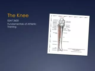
The Knee
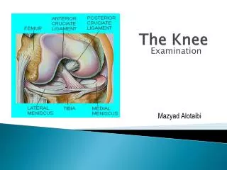
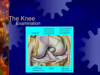
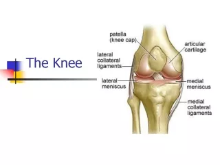
The Knee. Basic Anatomy. Basic Anatomy. Ligaments Anterior Cruciate Ligament (ACL) Prevents femur moving posteriorly during weight bearing Attaches anterior tibia to lateral femur Posterior Cruciate Ligament (PCL) Primary stabilizer & stronger than ACL


The Knee
E N D
Presentation Transcript
Basic Anatomy • Ligaments • Anterior Cruciate Ligament (ACL) • Prevents femur moving posteriorly during weight bearing • Attaches anterior tibia to lateral femur • Posterior Cruciate Ligament (PCL) • Primary stabilizer & stronger than ACL • Attaches posterior femur to anterior tibia • Medial Collateral Ligament (MCL) • Taut during knee flexion & relaxed in extension • Prevents knee from valgus and external rotating forces • Attaches medial femur & medial tibia • Lateral Collateral Ligament (LCL) • Taut during knee extension & relaxed in flexion • Attaches to the lateral femur & head of fibula
Meniscus • Medial meniscus: C-shaped fibrocartilage • Lateral meniscus: O-shaped fibrocartilage • Each meniscus is divided into 3 circumferential zones: • Outer zone = good vascular supply • Middle zone = minimal vascular supply • Innner zone = avascular
Prevention of Knee Injuries • Physical conditioning • Muscles surrounding knee joint must be strong and flexible • Focus on hamstring, erector spinae, groin, quadriceps, gastrocnemius flexibility • Rehabilitation and skill development • Strength, balance and technique - sport specific • Shoe Type • More and shorter cleats
MCL sprain Cause: • valgus force (lateral blow) or severe outward twist • Greater injury results from medial sprains vs. lateral sprains
S&S: • Grade I = few ligamentous fibers are torn and stretched; joint is stable, little/no swelling, possible joint stiffness & point tenderness, full ROM • Grade II = complete tear of deep cpsular ligament and partial tear of of MCL; slight laxity in full extension, no gross instability; no/little swelling; moderate to severe joint stiffness with inability to fully extend the knee; decrease PROM; medial pain • Grade III = complete loss of medial instability; swelling; immediate severe pain followed by dull ache; loss of motion because effusion and hamstring guarding Care: RICE; crutches; immobilzer; ROM exercises; rehabilitation; hinge brace (grade I, II)
LCL sprain Cause: • varus force & internal rotation of the tibia • Less prevalent than MCL
S&S: • Grade I = joint is stable, little/no swelling, possible joint stiffness & point tenderness, full ROM • Grade II = slight laxity in full extension, no gross instability; no/little swelling; moderate to severe joint stiffness with inability to fully extend the knee; decrease PROM; lateral pain • Grade III = complete loss of lateral instability; swelling; immediate severe pain followed by dull ache Care: RICE; crutches; immobilzer; ROM exercises;rehab; hinge brace; surgery controversial
ACL sprain Cause: foot planted, femur externally rotated S&S: hear/felt “pop”; immediate disability; rapid swelling; intense pain immediately then subsiding Care: RICE; crutches; immobilizer; surgery likely; 3-5 wks in brace; rehab 4-6 months
PCL sprain • Called the most important ligament in the knee • Provides central axis for rotation • Provides ~95% restraint of posterior displacement of tibia Cause: most at risk when knee is bent hyperflexed knee with full weight on anterior aspect of knee and foot plantarflexed; rotational force which may affect medial/lateral side of knee S&S: pop in back of knee; tenderness and swelling in back of knee (popliteal fossa); laxity of ligament Care: RICE; Grade 1 & 2 - non-op, rehab focusing on quad strengthening; rehabilitation; controversy over surgery
Meniscus injuries Cause: weight bearing combined with rotational stress while extending/flexing knee S&S: may or may not result in effusion gradually over 48-72 hrs; joint line pain and loss of motion; intermittent locking/giving way; pain with squatting Care: RICE; recovery depends on location of tear; not uncommon to return 6-14 days after surgery (menisectomy); repaired meniscus may require immobilization for 5-6 wks, longer recovery
Bursitis Cause: acute, chronic, recurrent; prepatellar and deep infrapatellar have highest incidence; prepatellar inflamed from continued kneeling or falling directly on knee; infrapatellar irritated from overuse of patellar tendon S&S: localized swelling; redness; increased temperature; pain due to swelling Care: eliminating cause; rest; swelling; compression; padding; NSAIDs; possible aspiration
Iliotibial band friction syndrome (Runners knee) • Overuse condition commonly in runners and cyclists who have genu varum and pronated feet • Irritation develops at the band’s insertion over the lateral femoral condyle causing friciton
Cause: overuse that can be attributed to malalignment and structural asymmetries of the foot and lower leg; S&S: tenderness along ITB; little/no swelling; pain increases with activity Care: stretch ITB; massage; foam roller: correction of foot and leg alignment problems; ice
Chrondromalacia • softening and deterioration of the articular cartilage on back of patella • Exact cause is unknown • Abnormal patellar tracking due to numerous factors Chondromalacia undergoes 3 stages: • Stage 1 = swelling and softening of articular cartilage • Stage 2 = fissuring of the softened articular cartilage • Stage 3 = deformation of the surface of the articular cartilage caused by fragmentation
Chondromalacia S&S: pain anterior knee while walking, running up/down stairs or squatting; recurrent swelling around kneecap; grating sensation when flex/ext knee Care: avoid irritating activities; isometric ex; NSAIDs; neoprene sleeve; possible sx
Patellar tendonitis (jumper’s knee) Cause: jumping, running, or kicking repetitively • Sudden or repetitive forceful extension of knee may begin an inflammatory process that will eventually lead to tendon degeneration S&S: tenderness; pain; • 3 stages: • Stage 1 - pain after activity • Stage 2 - pain during and after but functional • Stage 3 - pain during and prolonged after activity; may progress to constant Care: avoid irritating activities; RICE; patellar tendon brace; NSAIDs; transverse friction massage (TFM)
Osgood-Schlatter disease Cause: adolescent; repeated pull of patellar tendon at the tibial tubercle; bony callus forms and tubercle enlarges; usually resolves in late teen years S&S: repeated irritation causing swelling; hemorrhage, and gradual degeneration of tibial tubercle; severe pain when kneeling; jumping and running; point tenderness Care: stressful activities; ice before and after activity; isometric strengthening of quads and hams
Patellar dislocation Cause: quad muscle pulls in a straight line and patella pulls laterally creating a force that may sublux/dislocate patella S&S: complete loss of knee function; pain with swelling; obvious deformity; possible fracture Care: ice; referral with a dislocation - spontaneous reduction may occur if athlete is able to straighten knee
Osteochondritits Dissecans • A painful condition involving partial or complete separation of a piece of articular cartilage and subchondral bone • Exact cause is unknown • Possible causes = • direct or indirect trauma • skeletal or endocrine abnormalitites • prominent tibial spine or part of patella impinging on medial femoral condyle
S&S: achy knee, swells recurrently, may catch or lock, possible atrophy of quadriceps • Care: for children, rest and immobilization; may take as long as 1yr to resolve; surgery = drilling in the area to stimulate healing, pinning loose fragments, or bone grafting (Amare Stoudamire, Greg Oden)