

PKR -/- MEF
E N D
Presentation Transcript
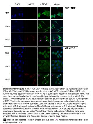
Supplementary figure 1. PKR null MEF cells are still capable of NF-kB nuclear translocation. IFA of WNV-induced NF-kB nuclear translocation in WT MEF cells and PKR null MEF cells. Twenty-four hrs post infection with WNV VLPs or 30min post treatment with 50ng/ml PMA, cell monolayers were fixed with 4% paraformaldehyde followed by permeablization with 0.1% triton-X-100 and blocked in 2% bovine serum albumin, 5% normal horse serum, 10mM glycine in PBS. The fixed monolayers were probed using the following monoclonal and polyclonal antibodies: anti-WNV MHIAF polyclonal, anti-NF-kB p65 (Santa Cruz), Alexa Fluor 488 goat anti-rabbit IgG (Invitrogen) and Alexa Fluor 568 goat anti-mouse IgG (Invitrogen). Following secondary antibody incubation, the cells were incubated with DAPI (500ng/ml) for nuclear counterstaining and mounted using VectaShield (Vector Laboratories). Stained cells were analyzed with a 1.0 Zeiss LSM 510 UV META Laser Scanning Confocal Microscope at the UTMB Infectious Disease and Toxicology Optical Imaging Core Facility. ( ) indicate translocated NFkB in antigen-positive cells, ( ) indicate untranslocated NFkB in antigen-positive cells DAPI a-WNV a-NFkB Merge PKR-/- MEF PMA Mock WNV WT MEF-G PMA Mock WNV