Download
1 / 44
480 likes | 567 Vues
Learn about epithelial tissues in animal structures, covering types, functions, and classifications. Explore simple squamous, cuboidal, columnar, and pseudostratified epithelia with glandular epithelium details. Discover the basement membrane, cell structures, and examples in organs.

E N D
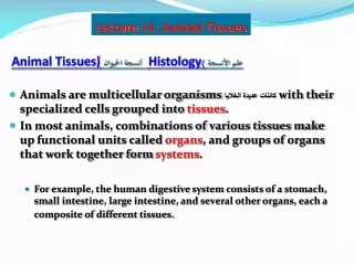
Animal Tissues By Fayez A. Elmabhouh Department of Biology
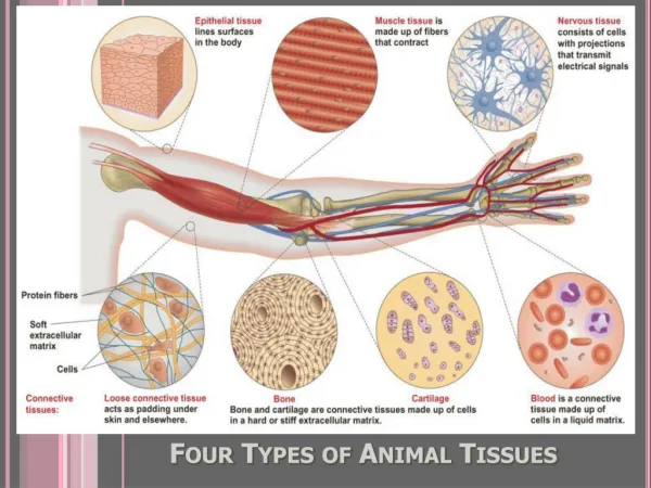
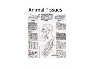
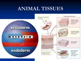
Introduction • Histology = study of tissues • Tissue = group of cells with similar structure and function • They are classified according to the shape of the cell, size, intracellular matrix. • Four types of human tissues: • Epithelial tissue • Connective tissue • Muscular tissue • Nervous tissue The organ can may consists of one (heart) or combination of these tissues (stomach, skeleton, skin)
Four Tissue Types: 1 2 3 4
Epithelial Tissues Characterized by: • Their closely connected cells. • Having very little intracellular substance. • Resting on a basement membrane. • Arise from the three germ layers. • No blood vessels enter between its cell but nerves do • Covers body surfaces and forms glands.
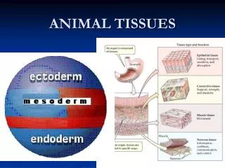
Embryonic Tissues – all adult tissues are derived from one of three embryonic tissues Ectoderm = “outside skin” gut Mesoderm = “middle skin” Cross section through embryo Endoderm = “inside skin” Animal embryo
Classification of Epithelium 1. Covering epithelia. 2. Glandular epithelia.
Covering epithelia • The primary function of this type of epithelia is protection. • Covering epithelia classified according to the arrangement of the cell into simple epithelia and stratified epithelia.
Simple epithelia • These tissues divided into four types according to the shape of cell and location of nucleus. • Consists of a single layer of cells that are in contact with the basement membrane.
1. Simple Squamous Epithelium: • The cells are flat with smooth edges. • They appear spindle-shaped in cross section • Each cell containing a nucleus in the widest area. • Found in the lining blood vessels, covering serosa
Simple Squamous Epithelium Simple Squamous Epithelium Bowman’s capsule
2. Simple Cuboidal Epithelium • The cells in this epithelium are square-shaped in cross section. • Have central and round nucleus. • Lining the kidney tubules and Follicles of thyroid gland
Simple Cuboidal Epithelium cells single layer of cube shaped cells; large nuclei
3. Simple Columnar Epithelium • Built of long pillar-shaped cells. • Containing an oval nucleus. • Ciliated: Bronchioles • Non Ciliated: lining the ileum
4. Pseudo-stratified Epithelium • Single layer of cells which rest on a basement membrane but do not all reach to the free surface of the epithelium. • Their nuclei found at different levels giving the a false stratified appearance. • Cells that reach the surface carry cilia at their free ends. • Lining the trachea.
Keratinized: the epithelium is covered with keratin layer which is formed by the dead squamous cells (horny layer) Skin. • Non- keratinized: esophagus
Keratinized: Skin • Epidermis (Stratified Squamous Epithelium) • Dermis
Transitional- stratified • Found in the urinary tract (urinary bladder) • Rest on a non clear and non wavy basement membrane. • Its superficial cells are cuboidal in shape, and may contain 2 nuclei. • The basal cell layer is formed of high cuboidal cell. • The intermediate layer are polyhydral cells. • Empty • Full, the cell change into to squamous cell
Glandular Epithelium • The cells are specialized in secretion and thus form glands. • Formed from collections of Epithelial cell • Glands with ducts are termed exocrine • Glands without ducts are termed endocrine
Exocrine Glands • Unicellular: formed of single cell present in the respiratory tract and intestinal tract
Multicellular gland • Made of many cells and each consist of a secretary portion and duct. • They are either tubular, alveolar in form and may be simple or compound.
Simple tubular glands • In the digestive glands, stomach, Large intestine
Simple alveolar gland • Form of a flask with a round secretary portion and a narrow tubular duct. • Mucous and sebaceous gland skin
Compound alveolar gland • Parotid gland