Embryonic Development: Medical Insights by Dr. Pooja Rajbhara
410 likes | 519 Vues
Learn about gestational age, organogenesis, layers of embryonic development, fetal milestones, and medical significance described by Dr. Pooja Rajbhara, a Senior Resident in Obstetrics & Gynecology.

Embryonic Development: Medical Insights by Dr. Pooja Rajbhara
E N D
Presentation Transcript
Development OF EMBRYO DR POOJA RAJBHARA SENIOR RESIDENT OBGY
GESTATIONAL AGE: • Gestational age or menstrual age is the time elapsed since the first day of the last menstrual period (LMP), a time that actually precedes conception. • Embryologists describe embryofetal development in terms of ovulation age, or the time in days or weeks from ovulation. • Another term is postconceptional age, which is nearly identical to ovulation age. • The period of gestation can also be divided into three units of approximately 14 weeks each. These three trimesters are important obstetrical milestones.
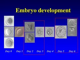
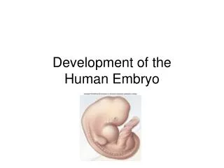
EMBRYONIC DEVELOPMENT: • The conceptus is termed an embryo at the beginning of the third week after ovulation and fertilization. • The embryonic period, during which time organogenesis takes place, lasts 8 weeks. • It begins the third week from the LMP through the eighth week. • The embryonic disc is well defined, and most pregnancy tests that measure human chorionic gonadotropin (hCG) become positive by this time. There are villous cores in which angioblastic chorionic mesoderm can be distinguished and a true intervillous space that contains maternal blood.
Ectodermal layer- CNS, PNS, epidermis of skin with its appendages, pituitary gland, chromaffin organs, salivary glands ,mucous lining of the nasal cavity, paranasal sinuses, roof of the mouth,etc. • Mesodermal layer- bones, cartilages, muscles, cvs, kidney, gonads, suprarenal, spleen, most of the genital tract, mesothelial lining of pericardial, pleural and peritoneal cavity,etc. • Endodermal layer- epithelial lining of git, liver, gallbladder, pancreas, epithelial lining of RS, most of the mucous membranes of urinary bladder and urethra, bulbourethral and greater vestibular glands, etc.
Cont: • During the 3 week, fetal blood vessels in the chorionic villi appear. • In the 4 week, a cardiovascular system has formed. Thereby, a true circulation is established both within the embryo and between the embryo and the chorionic villi. Partitioning of the primitive heart begins. Also in the fourth week, the neural plate forms, and it subsequently folds to form the neural tube.
. • By the end of the 5 menstrual week, the chorionic sac measures approximately 1 cm in diameter. The embryo is 3 mm long and can be measured sonographically. Arm and leg buds have developed, and the amnion is beginning to ensheathe the bodystalk, which thereafter becomes the umbilical cord.
. • At the end of the 6 week, the embryo measures approximately 9 mm long, and the neural tube has closed.Cardiac motion is almost always discernable sonographically. The neural tube has closed by the end of the sixth week
And by the end of the 8 week, the crown-rump length approximates 22 mm. Fingers and toes are present, and the arms bend at the elbows. The upper lip is complete, and the external ears form definitive elevations on either side of head.
Transition from the embryonic period to the fetal period occurs at 9 weeks after onset of the last menses. At this time, the fetus approximates 24 mm in length, most organ systems have developed, and the fetus enters a period of growth and maturation
12 weeks gestation: • Centers of ossification have appeared in most fetal bones, and the fingers and toes have become differentiated. • Skin and nails develop, and scattered rudiments of hair appear. • The external genitalia are beginning to show definitive signs of male or female gender. • The fetus begins to make spontaneous movements. 16weeks gestation: • Eye movements begin at 16 to 18 weeks, coinciding with midbrain maturation. • By 18 weeks in the female fetus, the uterus is formed and vaginal canalization begins. • By 20 weeks in the male, testicles start to descend.
20weeks gestation: • From this point onward, the fetus moves approximately every minute and is active 10 to 30 percent of the day. • Brown fat forms, and the fetal skin becomes less transparent. • Downy lanugo covers its entire body, and some scalp hair can be seen. • Cochlear function develops between 22 and 25 weeks, and its maturation continues for 6 months after delivery weeks gestation: 24 weeks gestation: • The fetus now weighs almost 700 g .The skin is characteristically wrinkled, and fat deposition begins. • The head is still comparatively large, and eyebrows and eyelashes are usually recognizable. • By 24 weeks, the secretory type II pneumocytes have initiated surfactant secretion The canalicular period of lung development, during which the bronchi and bronchioles enlarge and alveolar ducts develop, is nearly completed.
By 26 weeks, the eyes open. Nociceptors are present over all the body, and the neural pain system is developed .The fetal liver and spleen are important sites for hemopoiesis. • 28 weeks: The crown-rump length approximates 25 cm, and the fetus weighs about 1100 g. The thin skin is red and covered with vernix caseosa. Isolated eye blinking peaks at 28 weeks. The bone marrow becomes the major site of hemopoiesis. • 32 weeks of gestation:weight of about 1800 g. The skin surface is still red and wrinkled. • 36 weeks, the weight approximates 2800 g . Because of subcutaneous fat deposition, the body has become more rotund, and the previous wrinkled facies is now fuller.
40 weeks gestation: This is considered term, and the fetus is now fully developed. the average weight approximates 3500 g.
Fetal organogenesis:CENTRAL NERVOUS SYSTEM:BRAIN DEVELOPMENT
SPINAL CORD: • Whereas the superior two thirds of the neural tube give rise to the brain, the inferior third forms the spinal cord. • In the embryo, the spinal cord extends along the entire vertebral column length, but after that it lags behind vertebral growth. • Ossification of the entire sacrum is visible sonographically by approximately 21weeks • By 24 weeks, the spinal cord extends to S1, at birth to L3, and in the adult to L1. Spinal cord myelination begins at midgestation and continues through the first year of life. • Synaptic function is sufficiently developed by the eighth week to demonstrate flexion of the neck and trunk, During the third trimester, integration of nervous and muscular function proceeds rapidly
Cont: • Myelination of the ventral roots of the cerebrospinal nerves and brainstem begins at approximately 6 months, but most myelination progresses after birth. This lack of myelin and incomplete skull ossification permit fetal brain structure to be seen sonographically throughout gestation.
RESPIRATORY SYSTEM ANATOMICAL MATURATION: Within this framework, four essential lung development stages are described by . • First, the pseudoglandular stage entails growth of the intrasegmental bronchial tree between the 5th and 17th weeks. During this period, the lung looks microscopically like a gland. • Second, during the canalicular stage, from 16 to 25 weeks, the bronchial cartilage plates extend peripherally. Each terminal bronchio gives rise to several respiratory bronchioles, and each of these in turn divides into multiple saccular ducts.
. Third, the terminal sac stage begins after 25 weeks. During this stage, alveoli give rise to primitive pulmonary alveoli, that is, the terminal sacs. Simultaneously, an extracellular matrix develops from proximal to distal lung segments until term. • Finally, the alveolar stage begins during the late fetal period and continues well into childhood. An extensive capillary network is built, the lymph system forms, and type II pneumocytes begin to produce surfactant. At birth, only approximately 15 percent of the adult number of alveoli is present. Thus, the lung continues to grow, adding more alveoli for up to 8 years.
DIGESTIVE SYSTEM • After its embryogenic formation from the yolk sac as the primordial gut, the digestive system forms the intestines and various appendages. • The FOREGUT gives rise to the pharynx, lower respiratory system, esophagus, stomach, proximal duodenum, liver, pancreas, and biliary tree. • The MIDGUT gives rise to the distal duodenum, jejunum, ileum, cecum, appendix, and the right colon. • The HINDGUT develops into the left colon, rectum, and the superior portion of the anal canal. Numerous malformations develop in these structures from improper rotation, fixation, and partitioning.
Renal system: • Renal development involves interaction between pluripotential stem cells, undifferentiated mesenchymal cells, and epithelial components. • Two primitive urinary systems—the pronephros and the mesonephros—precede development of the metanephros, which forms the final kidney . • The pronephros involutes by 2 weeks, and the mesonephros produces urine at 5 weeks and degenerates by 11 to 12 weeks. • Failure of these two structures either to form or to regress may result in anomalous urinary system development.
. • . Between 9 and 12 weeks, the ureteric bud and the nephrogenicblastema interact to produce the metanephros. The kidney and ureter develop from intermediate mesoderm. • The bladder and urethra develop from the urogenital sinus. The bladder also develops in part from the allantois. • By week 14, the loop of Henle is functional and reabsorption occurs .New nephrons continue to be formed until 36 weeks. • In preterm neonates, their formation continues after birth.
Abnormalities of urinary tract: • Fetal hydronephrosis • Obstructive uropathy • Pelvic kidney • Horse shoe kidney • Polycystic kidney