Download
1 / 7
70 likes | 125 Vues
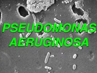
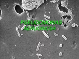
The study evaluates the impact of Mr. Trivedi’s biofield energy treatment on Pseudomonas aeruginosa (P. aeruginosa) to evaluate its phenotypic and genotypic characteristics.<br>

E N D
Trivedi et al.,J Trop Dis 2015, 4:1 http://dx.doi.org/10.4172/2329-891X.1000181 JournalofTropicalDiseases&PublicHealth Journal of Tropical Diseases ISSN: 2329-891X Research Article Research Article Open Access Open Access Antibiogram, Biochemical Reactions, and Genotypic Pattern of Biofield Treated Pseudomonas aeruginosa Mahendra Kumar Trivedi1, Alice Branton1, Dahryn Trivedi1, Gopal Nayak1, Mayank Gangwar2 and Snehasis Jana2* 1Trivedi Global Inc., 10624 S Eastern Avenue Suite A-969, Henderson, NV 89052, USA 2Trivedi Science Research Laboratory Pvt. Ltd., Hall-A, Chinar Mega Mall, Chinar Fortune City, Hoshangabad Rd., Bhopal- 462026 Madhya Pradesh, India Abstract Introduction: Complementary and alternative medicine such as biofield energy therapies are highly popular in biomedical health care. The study evaluates the impact of Mr. Trivedi’s biofield energy treatment on Pseudomonas aeruginosa (P. aeruginosa) to evaluate its phenotypic and genotypic characteristics. Methods: P. aeruginosa ATCC 10145 (American Type Culture Collection) was procured from Bangalore Genei, in sealed pack and divided into control and treated groups. Treated group was subjected to biofield treatment and analyzed for antibiogram, biochemical reactions, and biotype number using automated MicroScan Walk-Away® system on day 10. The treated sample was evaluated for DNA polymorphism by Random Amplified Polymorphic DNA (RAPD) and 16S rDNA sequencing to establish the phylogenetic relationship, the epidemiological relatedness and genetic characteristics. Results: Data showed altered sensitivity pattern in antibiotic cefotaxime from intermediate to decreased β-lactamases activity, with four-fold decreased minimum inhibitory concentration (MIC), i.e. 32 to ≤8 µg/mL as compared to control. Similarly, cefotetan and extended-spectrum-β-lactamases (ESBL-b Scrn) showed decrease in MIC values as compared to the control group. Nitrate reported for negative biochemical reaction i.e. positive (+) to negative (-) after biofield treatment on P. aeruginosa. The biotyping showed a change in biotype number (02063722) as compared to the control (02063726), without altering the microorganism. RAPD analysis showed an average range of 30 to 50% of polymorphism, while 16S rDNA sequencing analyzed treated sample as Pseudomonas aeruginosa (GenBank Accession Number: EU090892) with 99% identity of gene sequencing data. Conclusion: These results suggest that Mr. Trivedi’s unique biofield energy treatment on P. aeruginosa has an impact to alter the antimicrobial sensitivity pattern and MIC values, thus it can be used as an alternate integrative approach of energy medicine in near future. Keywords:Pseudomonas aeruginosa; Biofield energy treatment; Antibiogram; Polymorphism; RAPD; 16S rDNA analysis therapy approach creates the underlying selection pressure, and lead to develop resistance. A paradigm is now shifted with respect to the treatment of infectious diseases with antibiotics, where appropriate alternatives to antibiotics ought to be considered [10]. Several non-antibiotic approaches and prevention of infection including complementary and alternate medicines (CAM) are now preferred against P. aeruginosa [11]. CAM includes several energy medicines, among which biofield therapy (or healing modalities) is one of the approach, which was reported to have several benefits to enhance both physical and emotional human wellness [12]. Researchers are trying to investigate the relationship between biophysical, biochemical, and psycho-physiological mechanisms of disease and healing modalities. However, National Center for Complementary and Alternative Medicine (NCCAM) has funded to study the clinical application of biofield therapies in The Center for Frontier Medicine in Biofield Science (CFMBS), as a part of the National Institute of Abbreviations:NCCAM: National Center for Complementary and Alternative Medicine; NIH: National Institute of Health; CDC: Centers for Disease Control and Prevention; ATCC: American Type Culture Collection; DNA: Deoxyribonucleic acid; MIC: Minimum inhibitory concentration; MEGA: Molecular Evolutionary Genetics Analysis; NBPC 30: Negative Breakpoint Combo Panel 30; NCBI: National Center for Biotechnology Information; RAPD: Random Amplified Polymorphic DNA; OTUs: Operational Taxonomic Units; PCR: Polymerase Chain Reaction; RDP: Ribosomal Database Project Introduction The genus Pseudomonas is a Gram-negative, ubiquitous, rod- shaped, versatile opportunistic pathogen associated with nosocomial infections and other health related complications [1]. Some species are reported as pathogenic to plants [2] as well as opportunistic to animals or humans [3]. However, some species were reported to be used as bio-control agent, due to its plant growth promoter and pathogen suppressing function [4]. The increased prevalence of Pseudomonas aeruginosa in health care settings and its associated infections [5], restrain the effectiveness of antimicrobial therapy, which causes life threatening conditions [6]. It is one of the major pathogen associated with the hospital-acquired infections especially in intensive care unit [7]. The mechanism behind resistance pattern is due to acquisition of resistance genes (β-lactamases) or because of amino-glycoside modifying enzymes [8], or due to the chromosomal genes mutation involved against antimicrobials [9]. Unfortunately, the antimicrobial *Corresponding author: Snehasis Jana, Trivedi Science Research Laboratory Pvt. Ltd., Hall-A, Chinar Mega Mall, Chinar Fortune City, Hoshangabad Rd., Bhopal- 462026 Madhya Pradesh, India, Tel: 91-755-6660006; E-mail: publication@ trivedisrl.com Received September 10, 2015; Accepted September 30, 2015; Published S: October 07, 2015 Citation: Trivedi MK, Branton A, Trivedi D, Nayak G, Gangwar M, et al. (2015) Antibiogram, Biochemical Reactions, and Genotypic Pattern of Biofield Treated Pseudomonas aeruginosa. J Trop Dis 4: 181. doi:10.4172/2329-891X.1000181 Copyright: © 2015 Trivedi MK, et al.This is an open-access article distributed under the terms of the Creative Commons Attribution License, which permits unrestricted use, distribution, and reproduction in any medium, provided the original author and source are credited. J Trop Dis ISSN: 2329-891X JTD, an open access journal Volume 4 • Issue 1 • 1000181
Citation: Trivedi MK, Branton A, Trivedi D, Nayak G, Gangwar M, et al. (2015) Antibiogram, Biochemical Reactions, and Genotypic Pattern of Biofield Treated Pseudomonas aeruginosa. J Trop Dis 4: 181. doi:10.4172/2329-891X.1000181 Page 2 of 7 the standardized suspension of P. aeruginosa was pipetted into 25 mL of inoculum water using pluronic and inverted 8-10 times and inoculated, rehydrated, and then subjected to incubation for 16 hours at 35°C. Rehydration and inoculation were performed using the RENOK® system with inoculators-D (B1013-4). The detailed experimental procedures and conditions were followed as per the manufacturer’s instructions. Briefly, after inoculation and rehydration with a standardized suspension of P. aeruginosa, it was incubated at 35°C for 16 hours. The MIC and a qualitative susceptibility like susceptible (S), intermediate (I), and inducible β-lactamases (IB) were determined by observing the lowest antimicrobial concentration showing growth inhibition [24]. Biochemical studies The biochemical reactions of P. aeruginosa were determined by MicroScan Walk-Away® system, using photometric or fluorogenic reader. On the basis of nature of bacilli (Gram-negative or Gram- positive), computerized reports were generated using conventional panels, which utilizes the photometric reader. Before commencing the experiment, the NBPC 30 panel was first incubated and read on the MicroScan Walkaway system. After evaluating the experimental reading on the Walkaway system, the NBPC 30 panel was removed from system and recorded on the Biomic system within 1 hour. Instrument consist of a database associated with collective information, which was required to identify the microbes with respect to group, genera, or species of the family. Detailed experimental procedure was followed as per manufacturer-recommended instructions [24]. Biotype number The biotype number of P. aeruginosa was determined by MicroScan Walk-Away® processed panel data utilizing the data of biochemical reactions [24]. Random Amplified Polymorphic DNA (RAPD) analysis DNA fingerprinting was performed on three series of inoculum, using control and two treatment samples (A and B) prepared from P. aeruginosa. Two inoculums (A and B) were exposed with Mr. Trivedi’s biofield energy treatment. Further, the treated samples (A and B) were sub-cultured by taking 1% inoculum and inoculated to fresh 5 mL medium and labeled as treated A-1 and treated B-1. The samples were further incubated at 37°C with 160 rpm for 18 hour. Simultaneously, the cultures were spun down, and the genomic DNA was isolated for control and treated samples using Genomic DNA Prep Kit (Bangalore Genei, India). RAPD analysis was studied with all the samples of P. aeruginosa using standardised five RAPD primers, which were named as RBA8A, RBA13A, RBA20A, RBA23A, and RBA25A. The PCR mixture contained 2.5 μL of buffer, 4.0 mM of dNTP, 2.5 μM of primer, 5.0 μL of genomic DNA, 2U of Taq polymerase, 1.5 μL of MgCl2 and 9.5 μL of nuclease-free water in a total of 25 μL mixture. PCR amplification protocol followed with initial denaturation at 94ºC for 7 min, followed by 8 cycles of denaturation at 94ºC for 1 min, annealing at 35ºC for 1 min, and extension at 72ºC for 2 min; and 35 cycle of denaturation at 94ºC for 1 min, annealing at 38ºC for 1 min, and extension at 72ºC for 1.5 min; and the final extension at 72ºC for 7 min. The amplified PCR products (12 μL) from all samples (control and treated) were separated on 1% agarose gels at 75 volts, stained with ethidium bromide and visualized under UV illumination [25]. The percentage of polymorphism was calculated using following equation- Percent polymorphism = A/B×100; Health (NIH) [13]. Biofield is described as the endogenous energy field of the body, and defined as the electromagnetic field that permeates and surrounds living organisms [14]. Various internal physiological processes such as blood flow, brain, heart function, etc. that generate biofield. Biomagnetic fields around the human body can be measured using different techniques such as Kirlian photography, polycontrast interference photography (PIP) and resonance field imaging (RFI) [15]. Thus, it can be concluded that the human can harness the energy from the environment or universe and can transmit into any living or nonliving object(s) around the Universe. The objects always receive the energy and responding to the useful way that is called biofield energy treatment. Mr. Trivedi’s unique biofield treatment is also known as The Trivedi Effect®. It has been studied in the field of materials and ceramic sciences research [16,17], agricultural science research [18,19], biotechnology research [20], and altering the activity of pharmaceutical compounds [21], antimicrobials sensitivity of pathogenic microbes [22,23]. After evaluating the increasing use of CAM and failure of antimicrobial drug therapy against P. aeruginosa, the present study was designed to evaluate the impact of Mr. Trivedi’s biofield energy treatment on P. aeruginosa with respect to antibiogram analysis. The genotyping was performed to identify the strain differentiation and distinctive polymorphism using the polymerase chain reaction (PCR) technique of random amplified polymorphic DNA analysis (RAPD) and 16S rDNA sequencing. Materials and Methods P. aeruginosa ATCC 10145 (American Type Culture Collection) was procured from Bangalore Genei, in sealed pack, and stored as per the recommended storage conditions for further use. The antimicrobial susceptibility, biochemical reactions, and biotype number were evaluated using MicroScan Walk-Away® (Dade Behring Inc., West Sacramento, CA) using Negative Breakpoint Combo 30 (NBPC 30) panel. DNA fingerprinting (RAPD) and the 16S rDNA sequencing studies were carried out using Ultrapure Genomic DNA Prep Kit; Cat KT 83 (Bangalore Genei, India). All the tested antimicrobials, biochemicals, and reagents were procured from Sigma-Aldrich, India. Study design and biofield treatment P. aeruginosa strain was divided into two groups; one was kept as a control sample while the other was subjected to Mr. Trivedi’s unique biofield energy treatment and coded as treated group. The treated group was in sealed pack and handed over to Mr. Trivedi for biofield energy treatment under standard laboratory conditions. Mr. Trivedi provided the energy treatment through his energy transmission process that includes bioenergy emission of certain wavelength, which has the ability to do the changes in the microbes without touching the sample. Mr. Trivedi visited the laboratory individually over a period of treatment and for control experiments, nobody entered the experimental room during the treatment period. After treatment, control and treated groups were assessed on day 10 for antimicrobial susceptibility, minimum inhibitory concentration (MIC), biochemical reactions, biotype, and genotyping using RAPD and 16S rDNA sequencing analysis. Investigation of antimicrobial susceptibility assay Antimicrobial susceptibility assay of control and treated group of P. aeruginosa was carried out using MicroScan Walk-Away® using Negative Breakpoint Combo 30 (NBPC30) panel as per the manufacturer’s instructions. The panel was allowed to equilibrate to room temperature prior to rehydration. The test was carried out on MicroScan, which was miniaturized of the broth dilution susceptibility test. Briefly, 100 μL of J Trop Dis ISSN: 2329-891X JTD, an open access journal Volume 4 • Issue 1 • 1000181
Citation: Trivedi MK, Branton A, Trivedi D, Nayak G, Gangwar M, et al. (2015) Antibiogram, Biochemical Reactions, and Genotypic Pattern of Biofield Treated Pseudomonas aeruginosa. J Trop Dis 4: 181. doi:10.4172/2329-891X.1000181 Page 3 of 7 Where, A = number of polymorphic bands in treated sample; and B = number of polymorphic bands in control. Amplification and gene sequencing of 16S rDNA Gene sequencing was performed by isolating the genomic DNA and purified from a treated group of P. aeruginosa cells using genomic purification Kit, according to the manufacturer’s instructions. The PCR product was bi-directionally sequenced using the forward, reverse, and an internal primer. DNA 16S region amplification was performed using the primer set 16S forward and reverse primer [26]. 16S rDNA gene (~1.5 kb) was amplified by universal primers; forward primer (5ˊ-AGAGTTTGATCCTGGCTCAG-3ˊ) and reverse primer (3ˊ-ACGGTCATACCTTGTTACGACTT-5ˊ). carried out in a rapid cycler thermocontroller, with initial denaturation, annealing and extension temperature. Following amplification products were analyzed by gel electrophoresis at 100 V (in 1.0% agarose gel, 0.2 µg of ethidium bromide mL-1) in tris-acetate buffer (TAE), and visualized under UV light in a gel documentation unit (BioRad Laboratories, USA). The amplified fragment of PCR was purified from the agarose gel by DNA Gel Extraction Kit. The sequencing of amplified product was carried out on a commercial basis from Bangalore Genei, India. The obtained 16S rDNA sequences data were aligned and compared with the sequences, available in GenBank database of National Center for Biotechnology Information (NCBI) using the algorithm BLASTn program. The multiple sequence alignment/phylogenetic tree was constructed using MEGA 3.1 software using neighbor-joining method [27]. Results and Discussion Estimation of antimicrobial susceptibility The effect of biofield treatment on P. aeruginosa with respect to the antimicrobials susceptibility pattern and MIC are summarized in Tables 1 and 2, respectively. The data were analyzed and compared with respect to the control. The results showed an alteration of antibiogram in cefotaxime, as it was converted from intermediate (I) to inducible β-lactamases (IB), while no change in sensitivity pattern was recorded in rest of the tested antimicrobials in biofield treated P. aeruginosa as compared to the control. Similarly, MIC results of control and treated groups are summarized in Table 2. Antimicrobials such as cefotaxime, cefotetan, and ESBL-b Scrn were reported for altered MIC values in biofield treated P. aeruginosa with respect to the control. Biofield treated P. aeruginosa showed the four fold decrease in MIC values of cefotaxime (i.e. 32 to ≤8 µg/mL) as compared to the control microbe. Decreased MIC values was also reported in cefotetan (i.e. >32 to 32 µg/mL) and ESBL-b Scrn (i.e. >1 to ≤1 µg/mL) as compared to the control value. Biofield treated on P. aeruginosa did not show any alteration in MIC values in rest of the tested antimicrobials with respect to the control. The results of antimicrobial sensitivity assay showed an alteration of sensitivity pattern in biofield treated P. aeruginosa. Although antimicrobial resistance against various antimicrobials in clinical isolates, have been reported world-wide [28] which leads to a serious therapeutic threat. P. aeruginosa was reported to cause tissue damage in diabetes patients with foot ulcer [29]. According to Sivanmaliappan et al. the antibiogram pattern of P. aeruginosa revealed that cefotaxime and ciprofloxacin retained high activity as anti-pseudomonal drugs as compared to other antimicrobials. Hence, ciprofloxacin and cefotaxime were the best drugs of choice for diabetes patients with foot ulcers [29]. Its unique multiplicity towards the resistance mechanisms renders this microbe to treat by antibiotic therapy [30]. Experimental results showed an improve sensitivity and decrease MIC value of cefotaxime S. No. Antimicrobial Control Treated 1 Amikacin S S 2 Aztreonam IB IB 3 Cefepime S S 4 Cefotaxime I IB 5 Ceftazidime IB IB 6 Ceftriaxone IB IB 7 Ciprofloxacin S S 8 Gentamicin I I 9 Imipenem S S 10 Levofloxacin S S 11 Meropenem S S Amplification was 12 Piperacillin/tazobactam IB IB 13 Piperacillin IB IB 14 Ticarcillin/k-clavulanate IB IB 15 Tobramycin S S IB: Inducible β-lactamases; Resistant; I: Intermediate; S: Susceptible; ESBL: Suspected extended-spectrum β-lactamases; Antimicrobial susceptibility pattern in control and treated groups were evaluated using automated MicroScan Walk- Away® system using NBPC30 panel. Table 1: Effect of biofield treatment on multidrug resistant lab isolates of Pseudomonas aeruginosa to antimicrobial susceptibility. by four folds after biofield energy treatment on P. aeruginosa. Increase resistant of antimicrobials for P. aeruginosa has become a challenge for clinicians to select appropriate anti-pseudomonal antimicrobials. Antimicrobial sensitivity of cefotetan was also reported for broad spectrum of activity against Gram-negative microbes [31], but cefotetan will be highly active and shows synergistic action if taken in combination with other β-lactam antibiotics [32]. Biofield treatment result showed a slight decrease in MIC value of cefotetan as compared to control. Resistance mechanisms involved in P. aeruginosa includes aminoglycoside-modifying enzymes, efflux pumps, porin loss, and various target site modifications [33]. P. aeruginosa has been reported for acquired resistance by the production of plasmid mediated AmpC- β-lactamase, ESBL and metallo β-lactamase enzymes [34]. Biofield treatment has showed decreased MIC value of ESBL-b Scrn among the tested antimicrobials as compared to the control. The alterations might affect the β-lactamases production, which may lead to decrease the MIC, which is required to inhibit the growth of P. aeruginosa. Biofield energy treatment might act on the enzymatic or genetic level, which might affect the resistance mechanism, and lead to decrease the MIC value. Organism identification by biochemical reactions and biotype number The biochemical reactions of P. aeruginosa are presented in Table 3. Biofield energy treatment on P. aeruginosa showed an alteration of nitrate i.e. from positive (+) to negative (-) reaction as compared to the control. The rest of the thirty-two biochemicals did not show any change in the reaction pattern after biofield energy treatment. P. aeruginosa is a glucose non-fermenter, motile organism, shows oxidase positive, glucose negative, and Voges-Proskauer negative reactions as a characteristic feature. Biochemical reactions of control sample of P. aeruginosa were well supported with literature data [35]. P. aeruginosa was identified based on a variety of conventional biochemical characters and biotyping. After analyzing the results of the biochemical reactions, biotype number of P. aeruginosa was evaluated using automated Microscan system, which helps to identify the microorganism. In this experiment, biotyping was performed, and the results found a J Trop Dis ISSN: 2329-891X JTD, an open access journal Volume 4 • Issue 1 • 1000181
Citation: Trivedi MK, Branton A, Trivedi D, Nayak G, Gangwar M, et al. (2015) Antibiogram, Biochemical Reactions, and Genotypic Pattern of Biofield Treated Pseudomonas aeruginosa. J Trop Dis 4: 181. doi:10.4172/2329-891X.1000181 Page 4 of 7 S. No. Antimicrobial Control Treated S. No. Code Biochemical Control Treated 1 Amikacin ≤16 ≤16 1 ACE Acetamide + + 2 Amoxicillin/K-clavulanate >16/8 >16/8 2 ADO Adonitol - - 3 Ampicillin/sulbactam >16/8 >16/8 3 ARA Arabinose - - 4 Ampicillin >16 >16 4 ARG Arginine + + 5 Aztreonam ≤8 ≤8 5 CET Cetrimide + + 6 Cefazolin >16 >16 6 CF8 Cephalothin + + 7 Cefepime ≤8 ≤8 7 CIT Citrate + + 8 Cefotaxime 32 ≤8 8 CL4 Colistin - - 9 Cefotetan >32 32 9 ESC Esculin hydrolysis - - 10 Cefoxitin >16 >16 10 FD64 Nitrofurantoin + + 11 Ceftazidime ≤8 ≤8 11 GLU Glucose - - 12 Ceftriaxone ≤8 ≤8 12 H2S Hydrogen sulfide - - 13 Cefuroxime >16 >16 13 IND Indole - - 14 Cephalothin >16 >16 14 INO Inositol - - 15 Chloramphenicol >16 >16 15 K4 Kanamycin + + 16 Ciprofloxacin ≤1 ≤1 16 LYS Lysine - - 17 ESBL-a Scrn >4 >4 17 MAL Malonate + + 18 ESBL-b Scrn >1 ≤1 18 MEL Melibiose - - 19 Gentamicin 8 8 19 NIT Nitrate Oxidation- fermentation Galactosidase + - 20 Imipenem ≤4 ≤4 20 OF/G + + 21 Levofloxacin ≤2 ≤2 21 ONPG - - 22 Meropenem ≤4 ≤4 22 ORN Ornithine - - 23 Nitrofurantoin >64 >64 23 OXI Oxidase + + 24 Norfloxacin ≤4 ≤4 24 P4 Penicillin + + 25 Piperacillin/tazobactam ≤16 ≤16 25 RAF Raffinose - - 26 Piperacillin ≤16 ≤16 26 RHA Rhamnose - - 27 Tetracycline >8 >8 27 SOR Sorbitol - - 28 29 30 Ticarcillin/k-clavulanate Tobramycin Trimethoprim/sulfamethoxazole ≤16 ≤4 >2/38 ≤16 ≤4 >2/38 28 SUC Sucrose - - 29 TAR Tartrate Tryptophan Deaminase Tobramycin - - 30 TDA - - MIC values are presented in µg/mL; ESBL: Suspected extended-spectrum β-lactamases a, b screen; MIC values in control and treated groups were evaluated using automated MicroScan Walk-Away® system using NBPC30 panel. Table 2: Minimum inhibitory concentration (MIC) of tested antimicrobials against Pseudomonas aeruginosa 31 TO4 - - 32 URE Urea - - 33 VP Voges-Proskauer - - -: negative; +: positive; Biochemical reactions in control and treated groups were evaluated using automated MicroScan Walk-Away® system using NBPC30 panel. Table 3: Effect of biofield treatment on multidrug resistant lab isolates of Pseudomonas aeruginosa to the vital processes occurring in living organisms. change in biotype number (02063722) in treated group as compared to the control (02063726). Organism identified in both the groups was same as P. aeruginosa. Biotype number alteration was based on the biochemical characteristics. Our research group recently reported the alterations in biochemical reactions followed by the change in biotype number [22,23]. DNA fingerprinting by RAPD analysis Biofield energy treatment on P. aeruginosa was given to treated samples to determine the epidemiological relatedness and genetic characteristics. RAPD analysis was performed to study the correlation based on genetic similarity or mutations between the biofield treated samples and the control sample. RAPD analysis uses a short nucleotide primers, which were unrelated to known DNA sequences of the target organism [36]. DNA polymorphism can be efficiently detected using PCR primers and identify interstrain variations among species in treated samples. The degree of relatedness and genetic mapping can be correlated between similar or different treated sample species [37]. Random amplified polymorphic-DNA fragment patterns of P. aeruginosa control, and treated samples were generated using five RAPD primers and shown in Figure 1, with 100 base pair DNA ladder. The polymorphic bands in control and treated samples were marked by arrows. The RAPD patterns of treated samples showed some unique and dissimilar bands among control and treated samples. DNA polymorphism analyzed by RAPD analysis, a total number of bands, common, and unique bands are summarized in Table 4. The level of polymorphism in terms of percentage values between control and treated samples (A, A1, B, and B1) are presented in Table 5. The level of polymorphism was found with an average range of 30 to 50% in treated samples as compared to control, while 16 to 28% among treated samples of P. aeruginosa after the biofield treatment. The highest change in DNA sequence was observed in treated groups with RBA 23A primer as compared to control; however no change was found in treated group (control and A1, B, and B1; B and B1, A1 and B1) with RBA 20A primer as compared to control. RAPD also explain the relevant degree of genetic diversity. However, this technique has the potential to detected polymorphism throughout the entire genome. 16S rDNA genotyping The molecular PCR assay based on 16S rDNA amplification protocol using standard forward and reverse 16S universal primers have been commonly used as a taxonomic “gold standard” in identification J Trop Dis ISSN: 2329-891X JTD, an open access journal Volume 4 • Issue 1 • 1000181
Citation: Trivedi MK, Branton A, Trivedi D, Nayak G, Gangwar M, et al. (2015) Antibiogram, Biochemical Reactions, and Genotypic Pattern of Biofield Treated Pseudomonas aeruginosa. J Trop Dis 4: 181. doi:10.4172/2329-891X.1000181 Page 5 of 7 calculated and shown in Table 7. Phylogenetic tree of the partial 16S rDNA gene sequencing using MEGA 3.1 software by neighbor joining method are presented in Figure 2. The ten closely related bacterial species as Operational Taxonomic Units (OTUs) in order to investigate the phylogenetic relationship of P. aeruginosa among other ten other bacterial species were studied. There were 1500 base nucleotides of 16S rDNA gene sequences that were analyzed and multiple alignment were constructed using ClustalW in MEGA 3.1 software [27]. According to the data in Table 7, the lowest value of the genetic distance from P. aeruginosa was 0.006 base substitutions per site. All pairwise distance analysis was carried out using the p-distance method in MEGA 3.1. The proportion of remarked distance is also called p-distance and showed as the number of nucleotide distances site. Values in Table 7 were programmed into Figure 2 with optimal bootstrap consensus tree. In the phylogram, there were eleven OTUs. According to U.S. Department of Health and Human Services and Centers for Disease Control and Prevention (CDC), approximately 34% of adults in US used some kind of complementary health approach, among which healing therapies, therapeutic touch, and biofield energy treatment are some of the main approaches in energy medicine. Biofield therapies include Reiki, Qigong or non-contact therapeutic touch, which claims to reduce the pain, anxiety and promote the human wellbeing. Mechanism behind these healing modalities is based on modulating the patient-environment energy field along with healer’s biofield. Use of complementary and alternate medicine has several advantages instead of the current preferred treatment approach. National Institute of Health (NIH) defined this specialty as biofield in 1994, and it was accepted by US National Library of Medicine as a medical subject heading [39]. Bioelectromagnetic-based therapies and biofield therapies are energy therapies that use or manipulate the energy fields to promote health and healing. These energy therapies are well described under energy medicine by National Center for Complementary and Alternative Medicine (NIH/NCCAM) [40]. The study was designed todemonstrate the impact of Mr. Trivedi’s biofield energy treatment on The results suggest that Mr. Trivedi’s biofield energy treatment has an impact on P. aeruginosa strain, which resulted in altered antimicrobial sensitivity. Further, molecular methods were performed which showed the genetic Figure 1: Phylogenetic tree of the partial 16S rDNA gene sequencing of Pseudomonas aeruginosa biofield using MEGA 3.1 software by Neighbor joining method. Common bands in control and treated Unique band S. No. Primer Bands scored TSB- 1 Control TSA TSA-1 TSB 1 RBA 8A 16 6 0 1 3 0 0 2 RBA 13A 16 5 2 2 2 2 2 3 RBA 20A 12 6 1 2 0 0 0 4 RBA 23A 14 4 2 2 3 2 2 5 RBA 25A 7 4 1 2 0 2 0 TSA: treated sample A; TSA-1: treated sample A-1; TSB: treated sample B; TSB-1: treated sample B-1 Table 4: DNA polymorphism of treated biofield Pseudomonas aeruginosa analyzed by random amplified polymorphic DNA (RAPD) analysis. TSA and TSA-1 0% TSB and TSB-1 0% TSA and TSB 0% TSA-1 and TSB-1 34% C and TSA C and TSA-1 C and TSB C and TSB-1 Primer RBA 8A 41% 75% 41% 41% RBA 13A 30% 30% 30% 30% 40% 50% 0% 0% RBA 20A 44% 0% 0% 0% 25% 0% 44% 0% Alignment View Alignment results RBA 23A 66% 77% 55% 55% 77% 40% 11% 22% AN Sequence description RBA 15A Average polymorphism 71% 0% 28% 57% 0% 50% 14% 28% 7A 0.97 Sample studied 50% 36% 30% 36% 28% 28% 13% 16% Pseudomonas aeruginosa strain MML2212 Pseudomonas aeruginosa strain NK 2.1B-1 Pseudomonas aeruginosa isolate PAL106 EU344794 1.00 C: control; TSA: treated sample A; TSA-1: treated sample A-1; TSB: treated sample B; TSB-1: treated sample B-1 Table 5: Level of polymorphism between control and biofield energy treated Pseudomonas aeruginosa samples. EU352760 1.00 DQ464061 1.00 and determining the phylogenies of bacterial species [38]. 16S rDNA sequencing was performed in biofield treated P. aeruginosa to identify the other closely related species of treated sample. The alignment and comparison of the gene sequences were performed with the sequences stored in Gen Bank database available from NCBI using the algorithm BLASTn program. Based on nucleotides homology and phylogenetic analysis the microbe coded as sample 7A was closely detected as Pseudomonas aeruginosa strain R285 (GenBank Accession Number: EU090892) with 99% identity. The closest sequences of P. aeruginosa obtained from sequence alignment using NCBI GenBank and ribosomal database project (RDP) were presented in Table 6. Distance matrix based on nucleotide sequence homology (Using Kimura-2 Parameter) indicated nucleotide similarity and distance identities between sample ‘7A’ and other ten closest homologs microbe of P. aeruginosa was AB117953 1.00 Pseudomonas aeruginosa strain:WatG AJ249451 1.00 Pseudomonas aeruginosa strain AL98 AB126582 1.00 Pseudomonas aeruginosa EU090892 0.99 Pseudomonas aeruginosa strain R285 EU221381 1.00 Pseudomonas aeruginosa strain Y2P3 EF064786 1.00 Pseudomonas aeruginosa strain K3 AY548952 0.99 Pseudomonas aeruginosa strain Z5 Alignment results and sequence description has been obtained from the blast results of GenBank database of National Center for Biotechnology Information (NCBI) using the algorithm BLASTn program. AN: Accession number Table 6: The closest sequences of P. aeruginosa from sequence alignment using NCBI GenBank and ribosomal database project (RDP) J Trop Dis ISSN: 2329-891X JTD, an open access journal Volume 4 • Issue 1 • 1000181
Citation: Trivedi MK, Branton A, Trivedi D, Nayak G, Gangwar M, et al. (2015) Antibiogram, Biochemical Reactions, and Genotypic Pattern of Biofield Treated Pseudomonas aeruginosa. J Trop Dis 4: 181. doi:10.4172/2329-891X.1000181 Page 6 of 7 Distance Matrix AN 1 2 3 4 5 6 7 8 9 10 11 DQ464061 1 — 1 1 0.986 1 1 1 1 0.999 0.988 0.983 AB126582 2 0.000 — 1 0.986 1 1 1 1 0.999 0.988 0.983 EU352760 3 0.000 0.000 — 0.986 1 1 1 1 0.999 0.988 0.983 EU090892 4 0.014 0.014 0.014 — 0.986 0.986 0.986 0.986 0.985 0.999 0.994 AB117953 5 0.000 0.000 0.000 0.014 — 1 1 1 0.999 0.988 0.983 EU221381 6 0.000 0.000 0.000 0.014 0.000 — 1 1 0.999 0.988 0.983 EU344794 7 0.000 0.000 0.000 0.014 0.000 0.000 — 1 0.999 0.988 0.983 EF064786 8 0.000 0.000 0.000 0.014 0.000 0.000 0.000 — 0.999 0.988 0.983 AY548952 9 0.001 0.001 0.001 0.015 0.001 0.001 0.001 0.001 — 0.987 0.982 AJ249451 10 0.013 0.013 0.013 0.002 0.013 0.013 0.013 0.013 0.013 — 0.996 7A 11 0.017 0.017 0.017 0.006 0.017 0.017 0.017 0.017 0.018 0.004 — AN: Accession number; Total 1500 base nucleotide of 16S rDNA gene sequences were analyzed by multiple alignments using ClustalW program. Pairwise distance (lower left) and number of nucleotide difference (upper-right) for 16S forward and reverse primer was presented using Kimura-2 Parameters. Table 7: Distance matrix based on nucleotide sequence homology (Using Kimura-2 Parameter) indicates nucleotide similarity (above diagonal) and distance (below diagonal) identities between the studied sample ‘7A’ and ten other closest homologs microbe. participating members from PD Hinduja National Hospital and MRC, Mumbai, Microbiology Lab for conduction antimicrobial studies. Authors grateful to Bangalore Genei Private Limited, for conducting RAPD and 16S rDNA sequencing analysis. References 1. NJ Palleroni (1984) Genus I. Pseudomonas. Bergey’s manual of systematic bacteriology. Williams & Wilkins, Baltimore, MD, US. 2. Vos PD, Goor M, Gillis M, Ley JD (1985) Ribosomal ribonucleic acid cistron similarities of phytopathogenic Pseudomonas species. Int J Syst Evol Microbiol 35: 169-184. 3. Tyler SD, Strathdee CA, Rozee KR, Johnson WM (1995) Oligonucleotide primers designed to differentiate pathogenic pseudomonads on the basis of the sequencing of genes coding for 16S-23S rRNA internal transcribed spacers. Clin Diagn Lab Immunol 2: 448-453. 4. Keel C, Weller DM, Natsch A, Défago G, Cook RJ, et al. (1996) Conservation of the 2,4-diacetylphloroglucinol biosynthesis locus among fluorescent Pseudomonas strains from diverse geographic locations. Appl Environ Microbiol 62: 552-563. Figure 2: Random amplified polymorphic-DNA fragment patterns of Pseudomonas aeruginosa generated using five RAPD primers, RBA 8A, RBA 13A, RBA 20A, RBA 23A, and RBA 25A. 1: Control; 2: Treated A; 3: Treated A-1; 4: Treated B; 5: Treated B-1; M: 100 bp DNA Ladder. 5. Acar JF (1997) Consequences of bacterial resistance to antibiotics in medical practice. Clin Infect Dis 24 Suppl 1: S17-18. 6. Kerr KG, Snelling AM (2009) Pseudomonasaeruginosa: a formidable and ever- present adversary. J Hosp Infect 73: 338-344. alterations and similarities using RAPD and 16S rDNA sequencing methods. Conclusions Based on the study results, it was found that the cefotaxime sensitivity was changed, while decrease in MIC value was reported in case of cefotaxime (i.e. 32 to ≤ 8 µg/mL), cefotetan (i.e. >32 to 32 µg/mL) and ESBL-b Scrn (i.e. >1 to ≤1 µg/mL) as compared with the control. The biochemical reaction of nitrate was altered followed by a change in biotype number (02063722) in treated group as compared to the control biotype number (02063726). Using five RAPD markers, the sample was characterized and showed 30 to 50% interspecific polymorphic relationship with P. aeruginosa after biofield treatment as compared to the control. After biofield treatment, molecular analysis using 16S rDNA analysis showed that the sample was detected as Pseudomonas aeruginosa with 99% identity. So, it can be concluded that Mr. Trivedi’s biofield energy treatment on P. aeruginosa has an impact in altering the sensitivity of antimicrobials, which might be used as an alternative therapy (healing treatment) in future. 7. Rossolini GM, Mantengoli E (2005) Treatment and control of severe infections caused by multiresistant Pseudomonasaeruginosa. Clin Microbiol Infect 11 Suppl 4: 17-32. 8. Poole K (2005) Aminoglycoside resistance in Pseudomonas aeruginosa. Antimicrob Agents Chemother 49: 479-487. 9. Strateva T, Yordanov D (2009) Pseudomonas aeruginosa - a phenomenon of bacterial resistance. J Med Microbiol 58: 1133-1148. 10. Carson CF, Riley TV (2003) Non-antibiotic therapies for infectious diseases. Commun Dis Intell Q Rep 27 Suppl: S143-146. 11. Ulrey RK, Barksdale SM, Zhou W, van Hoek ML (2014) Cranberry proanthocyanidins have anti-biofilm properties against Pseudomonas aeruginosa. BMC Complement Altern Med 14: 499. 12. Movaffaghi Z, Farsi M (2009) Biofield therapies: biophysical basis and biological regulations? Complement Ther Clin Pract 15: 35-37. 13. Center for frontier medicine in biofield science (2007) University of Arizona and Institute for Frontier Science. 14. Jain S, Mills PJ (2010) Biofield therapies: helpful or full of hype? A best evidence synthesis. Int J Behav Med 17: 1-16. Acknowledgement 15. Prakash S, Chowdhury AR, Gupta A (2015) Monitoring the human health by measuring the biofield “aura”: An overview. IJAER 10: 27637-27641. This work was supported by Trivedi Science™, Trivedi Master Wellness™ and Trivedi Testimonials. Authors acknowledge the generosity and cooperation of all 16. Trivedi MK, Patil S, Nayak G, Jana S, Latiyal O (2015) Influence of biofield J Trop Dis ISSN: 2329-891X JTD, an open access journal Volume 4 • Issue 1 • 1000181
Citation: Trivedi MK, Branton A, Trivedi D, Nayak G, Gangwar M, et al. (2015) Antibiogram, Biochemical Reactions, and Genotypic Pattern of Biofield Treated Pseudomonas aeruginosa. J Trop Dis 4: 181. doi:10.4172/2329-891X.1000181 Page 7 of 7 treatment on physical, structural and spectral properties of boron nitride. J Material Sci Eng 4: 181. strains isolated from clinical sources. J Natl Med Assoc 96: 1065-1069. 29. Sivanmaliappan TS, Sevanan M (2011) Antimicrobial Susceptibility Patterns of Pseudomonas aeruginosa from Diabetes Patients with Foot Ulcers. Int J Microbiol 2011: 605195. 17. Trivedi MK, Nayak G, Patil S, Tallapragada RM, Latiyal O (2015) Studies of the atomic and crystalline characteristics of ceramic oxide nano powders after bio field treatment. Ind Eng Manage 4: 161. 30. de Man P, van Der Veeke E, Leemreijze M, van Leeuwen W, Vos G, et al. (2001) Enterobacter species in a pediatric hospital: horizontal transfer or selection in individual patients? J Infect Dis 184: 211-214. 18. Sances F, Flora E, Patil S, Spence A, Shinde V (2013) Impact of biofield treatment on ginseng and organic blueberry yield. Agrivita J Agric Sci 35: 22-29. 19. Lenssen AW (2013) Biofield and fungicide seed treatment influences on soybean productivity, seed quality and weed community. Agricultural Journal 8: 138-143. 31. Ward A, Richards DM (1985) Cefotetan. A review of its antibacterial activity, pharmacokinetic properties and therapeutic use. Drugs 30: 382-426. 32. Kubo M, Kato Y, Inamori Y (1985) Studies on the combination action of sisomicin, gentamicin and cefmenoxime, ceftizoxime, cefoperazone, cefotetan against Serratia marcescens and Pseudomonas aeruginosa. Jpn J Antibiot 38: 3487-3496. 20. Nayak G, Altekar N (2015) Effect of biofield treatment on plant growth and adaptation. J Environ Health Sci 1: 1-9. 21. Trivedi MK, Patil S, Shettigar H, Singh R, Jana S, et al. (2015) An impact of biofield treatment on spectroscopic characterization of pharmaceutical compounds. Mod Chem Appl 3: 159. 33. Zavascki AP, Carvalhaes CG, Picao RC, Gales AC (2010) Multidrug-resistant Pseudomonas aeruginosa and Acinetobacter baumannii: Resistance mechanisms and implications for therapy. Expert Rev Anti Infect Ther 8: 71-93. 22. Trivedi MK, Patil S, Shettigar H, Bairwa K, Jana S (2015) Phenotypic and biotypic characterization of Klebsiella oxytoca: An impact of biofield treatment. J Microb Biochem Technol 7: 203-206. 34. Aggarwal R, Chaudhary U, Bala K (2008) Detection of extended-spectrum beta-lactamase in Pseudomonas aeruginosa. Indian J Pathol Microbiol 51: 222-224. 23. Trivedi MK, Patil S, Shettigar H, Gangwar M, Jana S (2015) An effect of biofield treatment on Multidrug-resistant Burkholderia cepacia: A multihost pathogen. J Trop Dis 3: 167. 35. Arai T, Otake M, Enomoto S, Goto S, Kuwahara S (1970) Determination of Pseudomonas aeruginosa by biochemical test methods. II. Acylamidase test, a modified biochemical test for the identification of Pseudomonas aeruginosa. Jpn J Microbiol 14: 279-284. 24. Fader RC, Weaver E, Fossett R, Toyras M, Vanderlaan J, et al. (2013) Multilaboratory study of the biomic automated well-reading instrument versus MicroScan WalkAway for reading MicroScan antimicrobial susceptibility and identification panels. J Clin Microbiol 51: 1548-1554. 36. Williams JG, Kubelik AR, Livak KJ, Rafalski JA, Tingey SV (1990) DNA polymorphisms amplified by arbitrary primers are useful as genetic markers. Nucleic Acids Res 18: 6531-6535. 25. Welsh J, McClelland M (1990) Fingerprinting genomes using PCR with arbitrary primers. Nucleic Acids Res 18: 7213-7218. 37. Williams JG, Hanafey MK, Rafalski JA, Tingey SV (1993) Genetic analysis using random amplified polymorphic DNA markers. Methods Enzymol 218: 704-740. 26. Alm EW, Oerther DB, Larsen N, Stahl DA, Raskin L (1996) The oligonucleotide probe database. Appl Environ Microbiol 62: 3557-3559. 38. Woese CR (1987) Bacterial evolution. Microbiol Rev 51: 221-271. 27. Kumar S, Tamura K, Nei M (2004) MEGA3: Integrated software for Molecular Evolutionary Genetics Analysis and sequence alignment. Brief Bioinform 5: 150-163. 39. Rubik B (2002) The biofield hypothesis: its biophysical basis and role in medicine. J Altern Complement Med 8: 703-717. 28. Orrett FA (2004) Antimicrobial susceptibility survey of Pseudomonas aeruginosa 40. Clarke TC, Black LI, Stussman BJ, Barnes PM, Nahin RL (2015) Trends in the use of complementary health approaches among adults: United States, 2002- 2012. Natl Health Stat Report : 1-16. Submit your next manuscript and get advantages of OMICS Group submissions Unique features: • • • User friendly/feasible website-translation of your paper to 50 world’s leading languages Audio Version of published paper Digital articles to share and explore Special features: • • • • • • • • 400 Open Access Journals 30,000 editorial team 21 days rapid review process Quality and quick editorial, review and publication processing Indexing at PubMed (partial), Scopus, DOAJ, EBSCO, Index Copernicus and Google Scholar etc Sharing Option: Social Networking Enabled Authors, Reviewers and Editors rewarded with online Scientific Credits Better discount for your subsequent articles Citation: Trivedi MK, Branton A, Trivedi D, Nayak G, Gangwar M, et al. (2015) Antibiogram, Biochemical Reactions, and Genotypic Pattern of Biofield Treated Pseudomonas aeruginosa. J Trop Dis 4: 181. doi:10.4181/2329-891X.1000181 Submit your manuscript at: http://www.omicsonline.org/submission J Trop Dis ISSN: 2329-891X JTD, an open access journal Volume 4 • Issue 1 • 1000181