Download
1 / 11
110 likes | 509 Vues
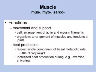
Muscle mus-, myo-, sarco-. Functions movement and support cell: arrangement of actin and myosin filaments organism: arrangement of muscles and tendons at joints heat production largest single component of basal metabolic rate 40% of body weight

E N D
Musclemus-, myo-, sarco- • Functions • movement and support • cell: arrangement of actin and myosin filaments • organism: arrangement of muscles and tendons at joints • heat production • largest single component of basal metabolic rate • 40% of body weight • increased heat production during, e.g., exercise, shivering
Muscle • Properties • excitable: responds electrically to external electrical, chemical or mechanical stimuli • e.g., generates action potentials • contractile (active) and extensible (passive) • due to actin and myosin interactions • elastic • Intracellular: due to cytoskeleton (e.g., titin) • Extracellular: due to extracellular matrix (e.g., collagen)
A skeletal muscle fiber is a syncytium. fusion mesodermal cells myoblasts muscle fiber Alberts, et al., Molecular Biology of the Cell. green = myosin muscle fiber = muscle cell
Skeletal Muscle Fibers Fig. 11.1
Levels of Organization Pattern of Bundles • myofilaments • actin and myosin • myofibril • myofiber • fascicle • anatomical muscle Fig. 10.1
Fascicle= little fasces http://www.classicsboy.50megs.com/cgibin/i/ Classics_Pictures/Man_Lictor_Bearing_Fasces.jpg http://reesbuilders.com/romanempire/images/guy.jpg
Alberts, et al., Molecular Biology of the Cell Extracellular Matrix • basal lamina similar to basement membrane • connective tissue with collagen fibers • contains blood vessels and nerves • continuous with tendons • integrates movements of individual muscle fibers • e.g., perimysium: surrounds fascicles • e.g., deep fascia: between or surrounding groups of anatomical muscles • loose connective tissue • superficial fascia = subcutaneous layer Fig. 10.1
Origin, Insertion, Belly Fig. 10.2
Fig. 10.4 Naming of Muscles shape location function Fig. 10.2
Arrangement of Fascicles pennate: force development and dexterity parallel: maximum shortening Fig. 10.3
Antagonistic and Synergistic Muscles Fig. 10.2 Hamstrings Fig. 10.4 Quadriceps femoris (“Quads”)