Download
1 / 49
490 likes | 724 Vues
The Autonomic Nervous System. Autonomic Nervous System. Assess Prof. Fawzia Al-Rouq Department of Physiology College of Medicine King Saud University. LECTUR (1). Functional Anatomy & Physiology of Autonomic NS. INTRODUCTION. THE NERVOUS SYSTEM. INTRODUCTION

E N D
The Autonomic Nervous System Autonomic Nervous System Assess Prof. Fawzia Al-Rouq Department of Physiology College of Medicine King Saud University
LECTUR (1) Functional Anatomy & Physiology of Autonomic NS
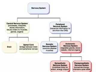
THE NERVOUS SYSTEM • INTRODUCTION • The nervous system monitors and controls almost every organ / system through a series of positive and negative feedback loops. • The Central Nervous System (CNS): Includes the brain and spinal cord. • The Peripheral Nervous System (PNS): Formed by neurons & their process present in all the regions of the body. • It consists of cranial nervesarises from the brain & spinal nerves arising from the spinal cord. • The peripheral NS is divided into • Somatic Nervous system • Autonomic nervous system
Anatomy and physiology of Autonomic Nervous System • At the end of this lectutre (1)the student should be able to:- • -appreciate the anatomy of symathetic& parasympathetic nervous system. • -explain physiological functions of Symathetic ¶sympathetic nerves in head&neck,chest,abdomen and pelvis
Basic anatomical difference between the motor pathways of the voluntary somatic nervous system (to skeletal muscles) and those of the autonomic nervous system • Somatic division: • Cell bodies of motor neurons reside in CNS (brain or spinal cord) • Their axons (sheathed in spinal nerves) extend all the way to their skeletal muscles • Autonomic system: chains of two motor neurons • 1st = preganglionic neuron (in brain or cord) • 2nd = gangionic neuron (cell body in ganglion outside CNS) • Slower because lightly or unmyelinated
Basic anatomical difference between the motor pathways of the voluntary somatic nervous system (to skeletal muscles) and those of the autonomic nervous system
The Autonomic Nervous System The Autonomic Nervous System1.Visceral sensory 2.Visceral motor
ANS is the subdivision of the peripheral nervous system that regulates body activities that are generally not under conscious control • Visceral motor innervates non-skeletal (non-somatic) muscles • Composed of a special group of neurons serving: • Cardiac muscle (the heart) • Smooth muscle (walls of viscera and blood vessels) • Internal organs • Skin
Axon of 1st (preganglionic) neuron leaves CNS to synapse with the 2nd (ganglionic) neuron • Axon of 2nd (ganglionic) neuron extends to the organ it serves this dorsal root ganglion is sensory
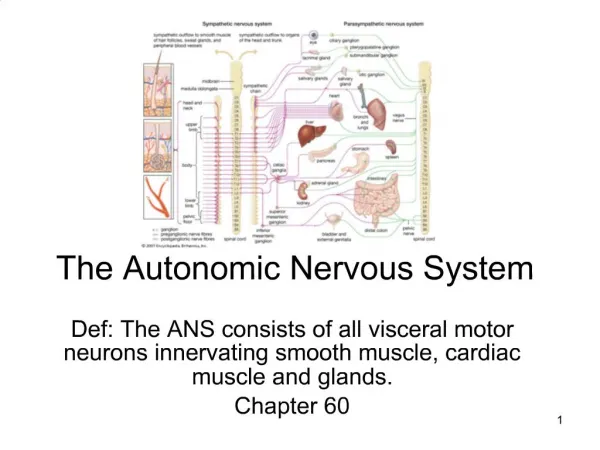
Overview of actions LOCATIONS OF AUTONOMIC GANGLIA • Sympathetic Ganglia: • Trunk (chain) ganglia near vertebral bodies • Prevertebral ganglia near large blood vessel in gut :celiac ,superior mesenteric & • inferior mesenteric • Parasympathetic Ganglia: • Terminal ganglia in the wall of organ
Spinal Cord Sympathetic Innervation of Visceral Targets • Short, lightly myelinated preganglionic neurons • Ganglia close to spinal cord • Long, unmyelinated postganglionic neurons
Parasympathetic Innervation of Visceral Targets • Ganglia close to or on target organs • Preganglionic neurons - long • Post ganglionic neurons - short
SYMPATHETIC & PARASYMPATHETIC NERVOUS SYSTEM ORIGIN Blue= Para symp; Red symp
Sympathetic - Origin • Thoracolumbar lateral horns of the spinal segments T1-L2. • Nerve fibers originate between T1 & L2
Parasympathetic - Origin • Craniosacral Cell bodies of the motor nuclei of the cranial nerves III, VII, IX and Xin the brain stem • Second, third and fourth [S2-S4] sacral segments of the spinal cord • Nerve fibers emerge from brain & • sacrum cranio-sacral outflow
PARASYMPATHETIC NERVOUS SYSTEM • The cranial nerves III, VII and IX affect the pupil and salivary gland secretion • Vagus nerve (X) carries fibres to the heart, lungs, stomach, upper intestine and ureter • The sacral fibres form pelvic plexuses which innervate the distal colon, rectum, bladder and reproductive organs.
SYMPATHETIC NERVOUS SYSTEM FUNCTIONS FEAR, FLIGHT OR FIGHT • The sympathetic system enables the body to be prepared for fear, flight or fight • Sympathetic responses include an increase in heart rate, blood pressure and cardiac output • Diversion of blood flow from the skin and splanchnic vessels to those supplying skeletal muscle • Increased pupil size, bronchiolar dilation, contraction of sphincters and metabolic changes such as the mobilisation of fat and glycogen.
FUNCTIONS OF SYMPATHETIC NERVOUS SYSTEM Bronchioles dilate, which allows for greater alveolar oxygen exchange. It increases heart rate and the contractility of cardiac cells (myocytes), thereby providing a mechanism for the enhanced blood flow to skeletal muscles. Sympathetic nerves dilate the pupil and relax the lens, allowing more light to enter the eye.
PARASYMPATHETIC NERVOUS SYSTEM FUNCTIONS • The parasympathetic nervous system has "rest and digest" activity. • In physiological terms, the parasympathetic system is concerned with conservation and restoration of energy, as it causes a reduction in heart rate and blood pressure, and facilitates digestion and absorption of nutrients, and consequently the excretion of waste products • The chemical transmitter at both pre and postganglionic synapses in the parasympathetic system is Acetylcholine (Ach).
THE AUTONOMIC NERVOUS SYSTEM
LECTUR (2) MECHANISM OF ACTIONS The neurotransmitters & receptors of Autonomic NS
OBJECTIVES • describe neurotransmitters that can release at pre and post ganglionic of Autonomic NS. • Describe Autonomic NS receptors.
Sympathetic Neurotransmitters • Preganglionic neurons - • Cholinergic = ( release acetylcholine ) • Postganglionic neurons: • release norepinepherine at target organs • ie. Adrenergic
Parasympathetic Neurotransmitters • Pre & Postganglionic neurons release acetylcholine = Cholinergic
ANS Neurotransmitters Classified as either cholinergic or adrenergic neurons based upon the neurotransmitter released • Cholinergic • Nor Adrenergic The neurons that are cholinergic are • Are pre ganglionic neurons • Anatomicallt para syampatheic post ganglionic neuron • Anatomical;lt syampatheic post ganglionic neuron ineervate sweet glands • Anatomically syampatheic neurons that end on blood vessels in skeletal muscles & produce vasodilatation • The remaining post ganglionic sympathetic neurons are nor adrenergic
ANS Neurotransmitters: Classified as either cholinergic or adrenergic neurons based upon the neurotransmitter released Cholinergic
Chemical or neural transmitter • All preganglionic fibers release acetylcholin (Ach). • All parasympatheticpostganglionic release Ach. • All sympathetic postganglionic release noradrenalin except sweat glands & bl vessels to skeletal muscles
Overview of actions RECEPTORS • The parasympathetic nervous system uses only acetylcholine (ACh) as its neurotransmitter. • The ACh acts on two types of receptors, the muscarinic and nicotonic choloinergic receptors. • Most transmissions occur in two stages: When stimulated, the preganglionic nerve releases ACh at the ganglion, which acts on nicotinic receptors of the postganglionic nerve. • The postganglionic nerve then releases ACh to stimulate the muscarinic receptors of the target organ. • The Sympathetic NS Acts on tow types of receptors :α and β.
Overview of actions TYPES OF MUSCARINIC RECEPTORS The three main types of muscarinic receptors: M1 muscarinic receptors: located in the neural system. M2 muscarinic receptors: located in the heart, and act to bring the heart back to normal after the actions of the sympathetic nervous system: slowing down the heart rate, reducing contractile forces of the atrial cardiac muscle, and reducing conduction velocity of the SA and AV node. Note, they have no effect on the contractile forces of the ventricular muscle.
Overview of actions TYPES OF MUSCARINIC RECEPTORS M3 muscarinic receptors: located at many places in the body, such as the smooth muscles of the blood vessels, as well as the lungs, which means that they cause vasoconstriction & bronchioconstriction and. They are also in the smooth muscles of the GIT, which help in increasing intestinal motility and dilating sphincters. M3 receptors are also located in many glands that help to stimulate secretion in salivary glands and other glands of the body.
Types of -adrenergic receptor • -adrenergic receptors are adrenergic receptors that respond to norepinephrine and to such blocking agents as phenoxybenzamine. • They are subdivided into two types: • 1, found in smooth muscle, heart, and liver, with effects including vasoconstriction, intestinal relaxation, uterine contraction and pupillary dilation, • 2, found in platelets, vascular smooth muscle, nerve termini, and pancreatic islets, with effects including platelet aggregation, vasoconstriction, and inhibition of norepinephrine release and of insulin secretion.
-receptor types • -adrenergic receptors respond particularly to epinephrine and to such blocking agents as propranolol. • There are three known types of beta receptor, designated β1, β2 and β3. • β1-Adrenergic receptors are located mainly in the heart. • β2-Adrenergic receptors are located mainly in the lungs, gastrointestinal tract, liver, uterus, vascular smooth muscle, and skeletal muscle. • β3-receptors are located in fat cells.
What do the receptors do?Activation of receptors leads to smooth muscle contractionActivation of 2 receptors leads to smooth muscle relaxationActivation of 1 receptors leads to smooth muscle contraction (especially in heart)