Download
1 / 18
271 likes | 1.35k Vues
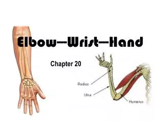
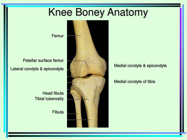
Knee Boney Anatomy. Medial condyle & epicondyle Medial condyle of tibia. Femur Patellar surface femur Lateral condyle & epicondyle Head fibula Tibial tuberosity Fibula. Medial Collateral Ligament. MCL Mechanism of Injury Valgus Stress. MCL Sprain.

E N D
Knee Boney Anatomy Medial condyle & epicondyle Medial condyle of tibia Femur Patellar surface femur Lateral condyle & epicondyle Head fibula Tibial tuberosity Fibula
MCL Mechanism of Injury Valgus Stress MCL Sprain 1st Degree 2nd Degree 3rd Degree
Valgus Stress Test Stresses MCL Valgus Stress at 0 - 5º Valgus Stress at 25 - 30 º
MOI of LCL Injury Varus Stress Test Stresses lateral structures Varus Stress Varus Stress at 0 º and 25 º to 30º of flexion
ACL Tear • Anterior instability • Mechanism • Deceleration injury • IR of femur with knee flexed and foot planted • Hyperextension of knee • Swelling • Pop at time of injury • Pain with • AROM • PROM • Anterior instability • Decreased strength • Giving way or buckling Signs and Symptoms
Anterior Drawer Test Grading Anterior InstabilityMedial viewRight knee • Stabilize Foot • Check for hamstrings relaxation • Thumbs either side patellar tendon • Apply anterior force • Grade amount of translation
Lachman’s Test • Better test than Anterior Drawer • Takes opposition of hamstrings out of play • Knee flexed 15 º - 30º • Stabilize femur • Apply anterior force to tibia
Pivot Shift Test • Gold standard test for ACL • Leg is externally rotated • Valgus force is applied as leg is flexed • Positive test indicated by clunk sensation
Posterior CruciateLigament Posterior cruciate
Posterior Sag Test • Posterior Cruciate vs Anterior Cruciate • Athlete supine • Both knees flexed 90’ • Observe laterally
Posterior Drawer Test • Athlete supine • Knee flexed 90’ • Foot neutral • Sit on foot to stabilize it • Posterior force applied at tibial plateau • Positive test indicates PCL injury PCL ACL
Medial and Lateral Meniscus Medial meniscus “C” shaped Lateral meniscus more circular shaped • Mechanism of Injury • Squat with rotation • Internal rotation of femur • on fixed tibia
Joint Space Orientation Lateral Meniscus Medial Meniscus Medial Joint Space Lateral Joint Space
Mc Murray Test • Flex knee fully • Palpate medial & lateral joint spaces with one hand • Rotate tibia opposite to femur as knee is extended • Palpable pop and/or pain indicate a positive test
Apley’s Compression Test External rotation of tibia tests medial meniscus Internal rotation of tibia tests lateral meniscus Apley’s Distraction Test Unloads the meniscus Stressess MCL and LCL