
INTRODUCTION
Amniotic fluid stem cells versus bone marrow mesenchymal stem cells for bone tissue engineering. 0 days. 7 days. 14 days. 21 days. 0 days. 7 days. 14 days. 21 days. 0 days. 7 days. 14 days. 21 days. 0 days. 7 days. 14 days. 21 days. MÁRCIA T. RODRIGUES*

INTRODUCTION
E N D
Presentation Transcript
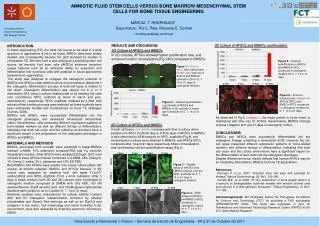
Amniotic fluid stem cells versus bone marrow mesenchymal stem cells for bone tissue engineering 0 days 7 days 14 days 21 days 0 days 7 days 14 days 21 days 0 days 7 days 14 days 21 days 0 days 7 days 14 days 21 days MÁRCIA T. RODRIGUES* Supervisors: Rui L. Reis, Manuela E. Gomes * mrodrigues@dep.uminho.pt hAFSCs hAFSCs hAFSCs hAFSCs hBMSCs hBMSCs hBMSCs hBMSCs 3D Culture of AFSCs and BMSCs (continuation) RESULTS AND DISCUSSION 2D Culture of AFSCs and BMSCs In 2D cultures, AFSCs showed higher proliferation rate, and enhanced matrix mineralization(Fig.1&2) compared to BMSCs. • INTRODUCTION • In tissue engineering (TE), the ideal cell source to be used in a wide spectrum of applications is yet to be found. BMSCs have been widely studied, and consequently became the gold standard for studies in orthopaedic TE. Amniotic fluid is also arising as a promising stem cell source, as amniotic fluid stem cells (AFSCs) evidence important unique features such as an extensive ability for expansion and differentiation into functional cells with potential in future approaches towards bone regeneration. • This study was designed to compare the osteogenic potential of BMSCs and AFSCs under distinct culture environments to determine if the osteogenic differentiation process of both cell types is related to cell origin. Osteogenic differentiation was carried out in 2 or 3 dimensions (3D) using a culture treated plate or by seeding the cells onto microfibrous SPCL scaffolds (a blend of starch and poly-caprolactone), respectively. SPCL scaffolds, obtained by a fiber melt extrusion/fiber bonding process were selected as these scaffolds have been extensively studied and characterized for bone TE strategies (Gomes. 2006). • BMSCs and AFSCs were successfully differentiated into the osteogenic phenotype, and developed mineralized extracellular matrix. Nevertheless, cells presented different expression patterns of bone-related markers as well as different timings of differentiation, indicating that both cell origin and the culturing environment have a significant impact in the progression of the osteogenic phenotype in AFSCs and BMSCs. • MATERIALS AND METHODS • BMSCs, purchased from Lonza®, were expanded in basal BMSCs medium: α-MEM, 10% embryonic screened-FBS and 1% penicillin /streptavidin solution, while AFSCs were isolated (DeCoppi. 2007) and cultured in basic AFSCs medium composed of α-MEM, 18% Chang B, 1% Chang C media, 2% L-glutamine and 15% ES-FBS. • Both BMSCs and AFSCs were seeded onto tissue culture plates (2D culture) at 30,000 cells/well. BMSCs and AFSCs behavior in 3D culture was assessed by seeding both cell types (1.2x10⁶ cells/scaffold) onto SPCL scaffolds (7mm x 4mm cylinders). After 3 days in basal medium, both 2D and 3D cultures were exchanged to osteogenic medium composed of DMEM with 10% FBS, 100 nMdexamethasone, 50µM ascorbic acid, and 10mM glycerol 2phosphate disodium salt hydrate for up to 3 weeks (0, 7, 14 or 21 days). • Retrieved samples were characterized for cellular viability (Calcein AM) and for osteogenic markers/matrix formation by alkaline phosphatase and Alizarin Red stainings as well as for RunX-2 and collagen I in the matrix. Cell morphology and matrix formation in 3D environment, were also assessed by scanning electronic microscopy (SEM). Figure 5 –Calcium quantification in AFSCs- and BMSCs-SPCL constructs in osteogenic medium for 0, 7, 14 or 21 days. Figure 1 – Viability assay withCalceinand Alizarin Red staining of AFSCs and BMSCs cultured in 2D for 0, 7, 14 or 21 days in osteogenic medium. • . Figure 6 – Immuno-fluorescence collagen I in AFSCs-SPCL and BMSCs-SPCL constructs in osteogenic medium for 0, 7, 14 or 21 days. Figure 2– Calcium quantification (g/ml/well) of AFSCs and BMSCs in 2D cultures in osteo-genic medium for 0, 7, 14 or 21 days. As observed in Fig.6, collagen I, the major protein in bone matrix is maintained with time only for AFSCs. Nevertheless, BMSCs strongly express collagen I at 0 and 21 days in osteogenic culture. CONCLUSIONS BMSCs and AFSCs were successfully differentiated into the osteogenic lineage, exhibiting a mineralized ECM. However the two cell types presented different expression patterns of bone-related markers, and different timings of differentiation, indicating that both cell origin and the culture environment have a significant impact on the differentiation of stem cells into the osteogenic phenotype. Despite differences found, results indicate that human AFSCs may be an interesting alternative to BMSCs for bone TE applications. References: DeCoppi P. et al. 2007. “Amniotic stem cell lines with potential for therapy.” Nature Biotechnology, 25, No1, 100-106. Gomes M.E. et al. 2006. “In vitro localization of bone growth factors in constructs of biodegradable scaffolds seeded with marrow stromal cells and cultured in a flow perfusion bioreactor.” Tissue Engineering, 12 No1, 177-188. Acknowledgements: MT Rodrigues thanks the Portuguese Foundation for Science and Technology (FCT) for providing a PhD scholarship (SFRH/BD/30745/ 2006). This study was supported, in part, by Telemedicine and Advanced Technology Research Center (TATRC) at the U.S. Army Medical Research. 3D Culture of AFSCs and BMSCs In both cell types, cell viability increases with time in culture when seeded onto SPCL scaffolds (Fig.3). A thick layer of AFSCs or BMSCs is also observed by SEM analysis on SPCL scaffolds (Fig.4), and mineralization nodules were detected in BMSCs- and AFSCs-SPCL constructs after 14 and 21 days respectively. Matrix mineralization was confirmed by calcium quantification assay (Fig.5). Figure 3 – Viability assay of hAFSCs and hMSCs cultured onto the SPCL scaffolds for 0, 7, 14 or 21 days in osteogenic medium. Figure 4 – SEMimages of AFSCs- and BMSCs-SPCL constructs in osteogenic culture for 0, 7, 14 or 21 days.


