Fungal Keratitis
1.75k likes | 4.75k Vues
Fungal Keratitis. Dr. Soujanya K MBBS, MS ( Ophthal ), DNB, FPRS Assistant Professor YMCH. Etiology. i . Filamentous fungi : Aspergillus (most common), Fusarium , Alternaria , Cephalosporium , Curvularia and Penicillium . ii. Yeasts: Candida and Cryptococcus.

Fungal Keratitis
E N D
Presentation Transcript
Fungal Keratitis Dr. Soujanya K MBBS, MS (Ophthal), DNB, FPRS Assistant Professor YMCH
Etiology • i. Filamentous fungi : • Aspergillus (most common), Fusarium, Alternaria, Cephalosporium, Curvulariaand Penicillium. • ii. Yeasts: Candida and Cryptococcus.
Modes of infection • i. Injury by vegetative material • ii. Injury by animal • iii. Patients who are immunosuppressedsystemically or locally
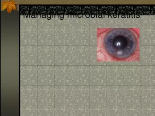
Clinical features • Symptoms : • are similar to the central bacterial corneal ulcer • less marked than the equal-sized bacterial ulcer • overall course is slow and torpid.
Symptoms Bacterial Fungal
Signs • Dryin appearance. • Feathery borders • An immune ring (Wesseley) : deposition of immune complexes and inflammatory cells around the ulcer. • Satellitelesions. • Hypopyon : thick and immobile, and is due to direct invasion into the anterior chamber of fungal hyphae enmeshed in thick exudates. • There is marked ciliaryand conjunctival congestion,
BacteralKeratitis Fungal Keratitis Symptoms are less Dry appearing, feathery margin May be present May be present Contains fungal hyphae, thick • Symptoms are more ( Pain, lid edema) • Round or oval ulcer in central part of cornea • Satelite lesions : Absent • Immune ring: Absent • Hypopyon : Sterile, mobile
Laboratory investigations • Wet KOH, • Calcofluor white, • Gram's and Giemsa- stained films for fungal hyphae and • Culture on Sabouraud's agar medium.
Treatment • 1. Topical antifungal eye drops should be used for a long period (6 to 8 weeks): • Natamycin (5%) eye drops • Fluconazole(0.2%) eye drops • Nystatin (3.5%) eye ointment.
2.Systemic antifungal drugs may be required for severe cases of fungal keratitis. • Tablet fluconazoleor ketoconazole may be given for 2-3 weeks.
Herpes simplex • Herpes zoster
HSV 1 HSV 2
Clinical reactivation • A variety of stressors such as • fever, • hormonal change, • ultraviolet radiation, • trauma
Herpes simplex – ocular manifestations • Blepharitis, • Conjunctivitis, • Keratitis • Iridocyclitis
Primary infection • No previous viral exposure, • Childhood • Subclinical or mild prodromal symptoms. • Blepharitis and follicular conjunctivitis :mild and self-limited. • Treatment: topical aciclovir ointment for the eye and/or cream for skin lesions.
Superficial Punctatekeratitis— • numerous minute whitish plaques , arranged in rows or groups. • Desquamate erosions heal rapidly leaving no opacity • Cornea: relatively insensitive.
The corneal involvement can be • Epithelial(dendritic or geographic keratitis), • Stromal(necrotizing and non-necrotizing stromalkeratitis) and • Endothelial
Dendriticulcers Pathognomonic of HSV keratitis
Floor of the ulcer stains with fluorescein and the virus-laden cells at the margin take up rose bengal.
fluorescein rose bengal
Branches of dendritic ulcer enlarge Steroid use Coalesce to form a large epithelial ulcer ( 'geographical' or 'amoeboid' configuration.)
Branches of dendritic ulcer enlarge Steroid use Coalesce to form a large epithelial ulcer ( 'geographical' or 'amoeboid' configuration.)
Branches of dendritic ulcer enlarge Steroid use Coalesce to form a large epithelial ulcer ( 'geographical' or 'amoeboid' configuration.)
Specific treatment • 1. Antiviral drugs are the first choice presently. • Aciclovir • Ganciclovir • Triflurothymidine • Vidarabine
2. Mechanical debridement : helps by removing the virus-laden cells.
Pathogenesis HSV
Low grade stromal inflammation and damage to the underlying endothelium Corneal oedema Disciformkeratitis