Download
1 / 50
500 likes | 743 Vues
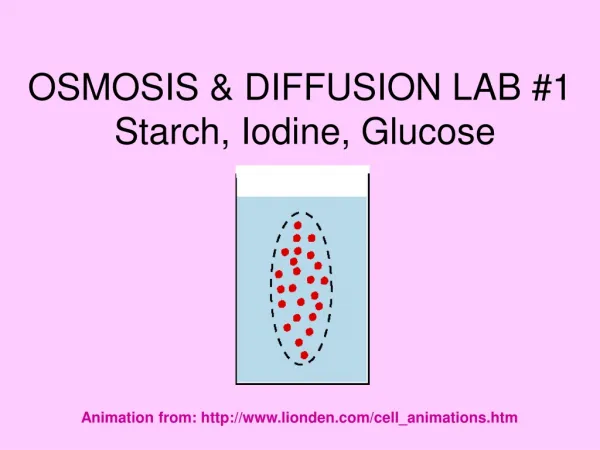
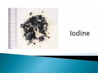
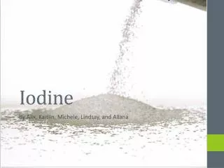
Iodine and Starch lab Show iodine with starch solution, sugar solution and distilled water Show dialysis tubing – it is semi-permeable Put starch solution in tubing, tie up and place in beaker of water. Put iodine into water – pictures would be helpful – Lab 1

E N D
Iodine and Starch lab Show iodine with starch solution, sugar solution and distilled water Show dialysis tubing – it is semi-permeable Put starch solution in tubing, tie up and place in beaker of water. Put iodine into water – pictures would be helpful – Lab 1 While we wait – go through the lab write-up sheet. We have done the section on data collection using tables. We will be looking at graphs to analysis our data. Then go through problem, hypothesis and variables. If time write up the procedure we did Check tubing, record data somehow Discuss/analysis results Now, what would happen if we put the starch in the beaker and the iodine in the tubing – lab 2
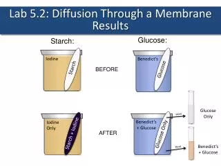
Requirements • Describe the results for lab 1. At least one picture is required. • Explain the results in paragraph form. This will help you figure out your hypothesis for lab 2. • State your hypothesis for the second procedure and give your reason why • List your independent and dependent variables and 3 good variables to control • Briefly describe the procedure you would do for lab 2. It can be in numbered form or paragraph but it must be reproducible. It should include a picture of the set-up • Explain why Step one was necessary
Cell Membrane Controls what enters and leaves the cell But HOW??
Its all in what it is made of and how it organizes the molecules
So, we know that the membrane is specially designed to help substances pass in and out. But how do things actually pass in and out???
http://www.wiley.com/legacy/college/boyer/0470003790/animations/membrane_transport/membrane_transport.htmhttp://www.wiley.com/legacy/college/boyer/0470003790/animations/membrane_transport/membrane_transport.htm
Good site for diffusion, osmosis etc http://www.biologycorner.com/bio1/notes_diffusion.html
Watch the cell membrane pull away from the cell wall Plant cells in a hypertonic solution
Doesn’t require energy Requires energy
Watch one celled organisms ooze in preparation for endocytosis
Take a lot of salt into your body and your metabolism very quickly goes into crisis. From every cell, water molecules rush off like so many volunteer firemen to try to dilute and carry off the sudden intake of salt. This leaves the cells dangerously short of the water they need to carry out their normal functions. They become, in a word, dehydrated. In extreme situations, dehydration will lead to seizures, unconsciousness and brain damage. Meanwhile, the overworked blood cells carry the salt to the kidneys, which eventually become overwhelmed and shut down. Without functioning kidneys you die. That is why we don’t drink sea water
We also know from doing a lab, that surface area matters. You have to touch a cell membrane (surface) before you can actually get into or out of a cell.
So if you get too large for your surface area, you have trouble moving substances in and out. You must divide instead!
PHASES OF MITOSIS *IPMAT • PROPHASE • METAPHASE • ANAPHASE • TELOPHASE
PROPHASE • 1st phase of mitosis • Chromosomes condense • Sister chromotids visible • Centrioles migrate to poles (animal cells) • Spindle fibers form and attach to centromeres • Nuclear membrane and nucleolus disappear
METAPHASE • META =MIDDLE • Chromosomes are pulled to the middle • Chromosomes line up on the Metaphase or Equatorial plate
ANAPHASE • Centromeres divide • Spindle fibers pull sets of CHROMATIDS towards opposite poles • CHROMATIDS separate to form 2 identical sets of daughter CHROMOSOMES • Ends when 2 sets are at opposite ends of the cell
TELOPHASE • Last phase of Mitosis (Reverse of Prophase) • Nuclear envelope (membrane) forms around each set of chromosomes. • Chromosomes uncoil Chromatin • Mitotic spindle fibers disassemble. • Membrane pinches in.
CYTOKINESIS • DIVISION OF THE CYTOPLASM • Animal Cells - Cell membrane pinches in until it meets and forms 2 cells • Plant Cells - Cell plate forms a double membrane, a new cell wall forms between them. WHY IS CYTOKINESIS DIFFERENT IN PLANTS AND ANIMALS?
Go to this site, click on onion cell activity in the left hand column. Read and go through until you get to the activity. http://www.biology.arizona.edu/ Interactive on mitosis http://biologyinmotion.com/cell_division/