NEUROMUSCULAR DISORDERS
NEUROMUSCULAR DISORDERS. COMMON NEUROMUSCULAR DISORDERS. Poliomyelitis Guillain Barre syndrome Muscular dystrophies Myasthenia gravis Spinal muscular atrophy (Werdnig Hoffman syndrome) Myopathies (Congenital & Acquired). Guillain Barre Syndrome (GBS).

NEUROMUSCULAR DISORDERS
E N D
Presentation Transcript
NEUROMUSCULAR DISORDERS TA OGUNLESI (FWACP)
COMMON NEUROMUSCULAR DISORDERS • Poliomyelitis • Guillain Barre syndrome • Muscular dystrophies • Myasthenia gravis • Spinal muscular atrophy (Werdnig Hoffman syndrome) • Myopathies (Congenital & Acquired) TA OGUNLESI (FWACP)
Guillain Barre Syndrome (GBS) • It is a cause of neuromuscular paralysis • It causes an acute, ascending, and progressive neuropathy characterized by weakness, paresthesias and hyporeflexia. • It is a post-infectious polyneuropathy that causes demyelination in mainly motor but sometimes also sensory nerves. • In severe cases, muscle weakness may lead to respiratory failure. Severe labile autonomic dysfunction also may occur. TA OGUNLESI (FWACP)
EPIDEMIOLOGY • GBS affects people of all ages but it is uncommon in children. • Sex incidence: Male-to-Female ratio is 1.5:1 • It is the most common neuromuscular disorder in the developed world unlike polio in the developing world. TA OGUNLESI (FWACP)
PATHOGENESIS • GBS is characterized by absent or profoundly delayed conduction in action nerve fibers. This aberrant conduction results from demyelination of peripheral nerves and spinal roots. Cranial nerves are rarely involved. • GBS results from an autoimmune response triggered by an antecedent viral infection. TA OGUNLESI (FWACP)
PATHOGENESIS • The autoimmune response appears to have both humoral and cell-mediated components. • The symptoms commonly result from immune-mediated injury to the myelin sheath. In a few cases, axonal damage results from a direct cellular immune attack on the axon itself. TA OGUNLESI (FWACP)
AETIOLOGY • Two thirds of patients have a preceding history of GI or respiratory infection in 1-3 weeks prior to the onset of weakness. • Infections like EBV, CMV, Chlamydia, HBV, Campylobacter, Mycoplasma, HIV • Vaccinations like Rabies, Streptococcus, Influenza. • Malignancies like lymphomas particularly Hodgkin disease. • Drugs like Captopril, Gold, Penicillamine. TA OGUNLESI (FWACP)
CLINICAL FEATURES • Weakness presenting in the ascending, symmetrical pattern starting from the lower extremities. Usually maximum in severity 2 weeks after the initial onset of symptoms. • Weakness may progress to inability or refusal to walk and later to flaccid tetraplegia. • Paresthesia with loss of sensation beginning at the toes and progressing upward and centrally. TA OGUNLESI (FWACP)
CLINICAL FEATURES • Pain over the lower back, buttocks and thighs. • Cranial nerves are involved in 45%-75% of cases. May present with facial weakness, dysphasia, drooling or dysarthrias. • In the Miller-Fisher variant, neuropathy begins with cranial nerve deficits. TA OGUNLESI (FWACP)
CLINICAL FEATURES • Autonomic dysfunctions like bradycardia or tachycardia, hypotension or hypertension, hypothermia or hyperthermia, anhidrosis, paralytic ileus and urinary retention. • Cranial nerve deficits: facial, bulbar palsies, muscles of mastication and ocular. • Papilledema may result from pseudotumor cerebri. • Hypotonia • Hyporeflexia or areflexia • Sensory loss • Respiratory embarrassment following diaphragmatic/ respiratory muscles involvement. TA OGUNLESI (FWACP)
CLINICAL COURSE • The clinical course is usually benign, and spontaneous recovery begins within 2–3 wk. • Most patients regain full muscular strength, although some are left with residual weakness. • Bulbar and respiratory muscle involvement may lead to death if treatment is delayed. TA OGUNLESI (FWACP)
CLINICAL COURSE • The tendon reflexes are usually the last function to recover. Improvement usually follows a gradient inverse to the direction of involvement, with recovery of bulbar function first and lower extremity weakness resolving last. TA OGUNLESI (FWACP)
DIAGNOSIS • Lumbar puncture and spinal fluid analysis • Most, but not all, patients have an elevated level of CSF protein (>400 mg/L), with no elevation in CSF cell counts (Cyto-albumin dissociation). • CSF protein may be normal very early in the disease. • Serology: Four-fold rise in serum antibodies against microbes • Antibody screen • Antibodies to peripheral and central nerves may be present. TA OGUNLESI (FWACP)
DIAGNOSIS • Electromyogram shows evidence of acute denervation of muscle. Serum creatine phosphokinase (CK) level may be mildly elevated or normal. Muscle biopsy is not usually required for diagnosis • Lung Function Tests (Forced vital capacity in cases of respiratory muscle involvement) • Nerve conduction studies shows slowing 2-3 weeks after the onset of illness. • Electrocardiogram may show disorders of conduction and rhythm. TA OGUNLESI (FWACP)
DIFFERENTIAL DIAGNOSIS • Poliomyelitis • Hypokalaemia • Botulism • Cauda equina syndrome • Diptheria TA OGUNLESI (FWACP)
DIFFERENTIAL DIAGNOSIS • Myasthenia gravis • Spinal cord injuriess • Transverse myelitis • Organophosphate poisoning • Hereditary neuropathies • Porphyria TA OGUNLESI (FWACP)
COMPLICATIONS • Respiratory failure • Hypotension or hypertension • Thromboembolism (DVT) • Hypostatic pneumonia • Decubitus ulcers • Cardiac arrhythmia • Ileus • Aspiration • Urinary retention TA OGUNLESI (FWACP)
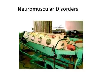
MANAGEMENT • ABCs, IVF, oxygen, and assisted ventilation may be indicated. • Adequate nursing care to prevent pressure sores • Intubation should be performed in the presence of hypoxia, rapidly declining respiratory function, poor or weak cough, and suspected aspiration. Typically, intubation is indicated whenever the FVC is less than 15 mL/kg. • Physiotherapy TA OGUNLESI (FWACP)
MANAGEMENT • Patients who have, or are suspected of having, GBS should be monitored closely for changes in blood pressure, heart rate, and other arrhythmias. • Treatment rarely is needed for tachycardia. • Atropine is recommended for symptomatic bradycardia. • Hypertension may be treated with short-acting agents like beta-blocker or nitroprusside. • Hypotension of dysautonomia usually responds to intravenous fluids and supine positioning. TA OGUNLESI (FWACP)
MANAGEMENT • Only plasma exchange therapy and IV immune serum globulin have proven effective. Both therapies have been shown to shorten recovery time by as much as 50%. • Deep vein thrombosis (DVT) prophylaxis with gradient compression hose and subcutaneous low molecular weight heparin (LMWH) may cause a dramatic reduction in the incidence of venous thromboembolism, one of the major sequela of extremity paralysis. • Prednisolone 2mg/kg/day is ineffective by itself. It may be given alongside plasmapheresis. TA OGUNLESI (FWACP)
PROGNOSIS • Peak weakness occurs in 10-14 days with recovery in weeks to months. • Full recovery occurs in 50-95% of cases. • Residual neurologic sequelae of varying degree may be observed in 10%-40% of cases. • Mortality rates range from 5%-10%. • Most mortality is due to severe autonomic instability or from the complications of prolonged intubation and paralysis. • Recurrence is rare but has been reported in 2%-5% of cases. TA OGUNLESI (FWACP)
MUSCULAR DYSTROPHIES • Muscular dystrophy is distinguished from all other neuromuscular diseases by four obligatory criteria: (1) It is a primary myopathy; (2) it has a genetic basis; (3) the course is progressive; (4) degeneration and death of muscle fibers occur at some stage in the disease. • Common examples are Duchenne, Bekers and Limb – Girdle muscular dystrophies. TA OGUNLESI (FWACP)
DUCHENNE MUSCULAR DYSTROPHY • Duchenne muscular dystrophy is the most common hereditary neuromuscular disease affecting all races. • It is inherited as an X-linked recessive trait. • Becker muscular dystrophy is a mild form of Duchenne dystrophy. • It is due to deficiency of dystrophin & characterized by excessive necrosis of the muscle fibres. TA OGUNLESI (FWACP)
CLINICAL FEATURES • Boys are exclusively affected. • Infants are rarely symptomatic at birth or in early infancy. • Early gross motor skills may be normal or slightly delayed. • Distinctive facies resulting from facial weakness is a late feature. • Hip girdle weakness may be seen as early as the 2nd year. • An early Gowers sign is often evident by age 3yr and is fully expressed by age 5 or 6yr. A Trendelenburg gait, or hip waddle, appears at this time. TA OGUNLESI (FWACP)
CLINICAL FEATURES • The progression of weakness continues into the 2nd decade. The function of distal muscles is usually relatively well enough preserved. • Respiratory muscle involvement is expressed as a weak and ineffective cough, frequent pulmonary infections, and decreasing respiratory reserve. • Pharyngeal weakness may lead to episodes of aspiration, nasal regurgitation of liquids and nasal speech. TA OGUNLESI (FWACP)
CLINICAL FEATURES • Incontinence due to anal and urethral sphincter weakness is a very late event. • Contractures most often involve the ankles, knees, hips, and elbows. • Scoliosis and lordosis are common. • Enlargement of the calves (pseudohypertrophy) and wasting of thigh muscles is a classic feature. The muscles of the tongue are also hypertrophied but without fasciculations. TA OGUNLESI (FWACP)
CLINICAL FEATURES • The deep tendon reflexes are gradually lost. • Cardiomyopathy is a constant feature of this disease. • Intellectual impairment occurs in all patients, although only 20–30% have significant mental retardation. • Epilepsy is uncommon but still slightly more common than in the general pediatric population. TA OGUNLESI (FWACP)
CLINICAL FEATURES • Muscle fibres are gradually replaced with fibrotic tissues and patients become wheel-chair bound by early adolescence. • Death from respiratory failure and CCF usually occur by about 18 years. TA OGUNLESI (FWACP)
DIAGNOSIS • The serum Creatine Kinase level is consistently greatly elevated even at birth. • Cardiac assessment by echocardiography, electrocardiogram (ECG) and Chest X-Ray is essential and should be repeated periodically. • Electromyogram (EMG) shows characteristic non-specific myopathic features. TA OGUNLESI (FWACP)
DIAGNOSIS • No evidence of denervation is found. Motor and sensory nerve conduction velocities are normal. • Muscle biopsy is diagnostic. • Immunohistochemistry to demonstrate deficient or defective dystrophin. TA OGUNLESI (FWACP)
MANAGEMENT • Physiotherapy with the use of orthotics • Anti-CCF regime with digoxin and diuretics • Antibiotics when chest infections occur. • Steroid trial. • Genetic engineering to produce efficient dystrophin. TA OGUNLESI (FWACP)
MYASTHENIA GRAVIS • Myasthenia gravis (MG) is an acquired autoimmune mediated neuromuscular blockade characterized clinically by weakness of skeletal muscles and fatigability on exertion. • EPIDEMIOLOGY: In the US, incidence is 2/1,000,000. Incidence in Nigeria is unknown. Sex incidence: Female:Male = 6:4 Age incidence: MG presents at any age but it is uncommon in childhood. It occurs in infancy either as CONGENITAL MG or TRANSIENT NEONATAL MG. TA OGUNLESI (FWACP)
CLINICAL TYPES • Acquired: Usually an autoimmune disease • Congenital: Occurs in newborn infants of non-myasthenic mothers. No spontaneous remission; persists with age. • Transient: Occurs in newborn infants of myasthenic mothers who acquire destructive antibodies transplacentally. Improve when antibodies disappear. • Familial: Rare; autosomal recessive; not associated with plasma AChR antibodies. TA OGUNLESI (FWACP)
NEUROMUSCULAR JUNCTION • The nerve terminal of the motor nerve enlarges at its end, which is called the terminal bulb. It lies within a groove or indentation along the muscle fiber. The presynaptic membrane (nerve membrane), postsynaptic membrane (muscle membrane), and synaptic cleft (space between the 2 membranes) together constitute the NMJ. TA OGUNLESI (FWACP)
PATHOPHYSIOLOGY • The presynaptic terminal contains vesicles filled with acetylcholine (ACh). On arrival of a nerve action potential, the contents of these vesicles are released into the synaptic cleft in a calcium-dependent manner. The released ACh molecules diffuse across the synapse and bind to the AChRs on the postsynaptic membrane. • ACh molecules are hydrolyzed by the enzyme acetylcholinesterase (AChE), which is abundantly present at the neuromuscular junction. TA OGUNLESI (FWACP)
PATHOPHYSIOLOGY • The antibodies in MG are directed toward the acetylcholine receptor (AChR) at the NMJ resulting in a decrease in the number of available ACh receptors at the NMJ of skeletal muscles. • The release of acetylcholine (ACh) into the synaptic cleft by the axonal terminal is normal, but the postsynaptic muscle membrane or motor end plate is less responsive because of decrease in the number of AChR. • The lack of response to ACh results in progressive muscular weakness. TA OGUNLESI (FWACP)
CLINICAL FEATURES • It is characterized by fluctuating muscle weakness usually increased by exertion. • Ptosis and diplopia. • Myopathic-like limb weakness (proximal worse than distal) • Weakness progress from mild to more severe disease over weeks to months. It tends to spread from the ocular to facial to bulbar muscles and then to truncal and limb muscles. TA OGUNLESI (FWACP)
CLINICAL FEATURES • Weakness can be present in a variety of different muscles and is usually proximal and symmetric. • Sensory examination and deep tendon reflexes are normal. TA OGUNLESI (FWACP)
CLINICAL FEATURES • Bulbar muscle weakness (nasal speech, food regurgitation, slow chewing, jaw hanging open, dysphagia) • Respiratory muscle weakness may produce acute respiratory failure which often results in death. TA OGUNLESI (FWACP)
CLINICAL FEATURES • Evidence of other coexisting autoimmune diseases: • Haemolytic anaemia • Grave’s disease • SLE • JRA • Scleroderma • No fasciculations, sensory loss or myalgia TA OGUNLESI (FWACP)
CLINICAL FEATURES • Weakness typically worsens with prolonged muscle activity: • Ptosis increases progressively when the patient tries to sustain an upward gaze for 30–90sec. • Repetitive opening and closing of the fists produces rapid fatigue of hand muscles • Inability to elevate the arms for more than 1–2min because of fatigue of the deltoids. TA OGUNLESI (FWACP)
LABORATORY FINDINGS • Electromyogram (EMG) shows a decremental response to repetitive nerve stimulation. This pattern is reversed after a cholinesterase inhibitor is administered. Anti-ACh antibodies should be assayed in the plasma but are inconsistently demonstrated. Antinuclear antibodies may be present. • Serum creatine phosphokinase (CK) level is normal. • ECG findings are usually normal. TA OGUNLESI (FWACP)
LABORATORY FINDINGS • Chest X-Ray reveals an enlarged thymus, but the hypertrophy is not usually a thymoma. It may be further defined by tomography or by CT imaging of the anterior mediastinum. • Muscle biopsy is not very helpful. • TENSILON test: A short-acting cholinesterase inhibitor, usually edrophonium chloride (0.2mg/kg IV) or neostigmine (0.4mg/kg IM) is administered. Within a few seconds, the ptosis and ophthalmoplegia improve, and fatigability of other muscles is greatly decreased. The effects last only 1–2min. TA OGUNLESI (FWACP)
TREATMENT • Some patients with mild myasthenia gravis require no treatment. • Cholinesterase-inhibiting drugs like neostigmine methylsulfate (0.04mg/kg IM q 4-hourly OR 0.4mg/kg orally q 4-hourly) are the primary therapeutic agents. Pyridostigmine may also be used in higher doses. • Long-term steroid treatment with prednisolone (0.2mg/kg/day) may be effective. TA OGUNLESI (FWACP)
TREATMENT • Thymectomy is most effective in patients with high titers of anti-Ach-receptor antibodies. It is ineffective in congenital and familial forms of myasthenia gravis. • Plasmapheresis is effective in children who do not respond to steroids. • Intravenous immunoglobulin (IVIG) is most useful when combined with IVIG. • Neonates with transient maternally transmitted myasthenia gravis require cholinesterase inhibitors for only a few days. TA OGUNLESI (FWACP)
COMPLICATIONS • Inter-current infections & antibiotics like gentamicin may precipitate myasthenia crisis. • Acute respiratory failure. • Aspiration pneumonitis. • Malnutrition from dysphagia. TA OGUNLESI (FWACP)