Figure 32.1
Figure 32.1. Zygote. Cleavage. Figure 32.2-1. Eight-cell stage. Zygote. Cleavage. Blastocoel. Figure 32.2-2. Cleavage. Eight-cell stage. Cross section of blastula. Blastula. Zygote. Cleavage. Blastocoel. Figure 32.2-3. Cleavage. Gastrulation. Eight-cell stage.
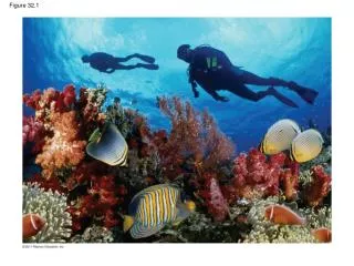

Figure 32.1
E N D
Presentation Transcript
Zygote Cleavage Figure 32.2-1 Eight-cellstage
Zygote Cleavage Blastocoel Figure 32.2-2 Cleavage Eight-cellstage Cross sectionof blastula Blastula
Zygote Cleavage Blastocoel Figure 32.2-3 Cleavage Gastrulation Eight-cellstage Cross sectionof blastula Blastula Blastocoel Endoderm Ectoderm Archenteron Cross sectionof gastrula Blastopore
Individualchoanoflagellate Figure 32.3 Choanoflagellates OTHEREUKARYOTES Sponges Animals Collar cell(choanocyte) Other animals
2 1 3 4 RESULTS Early stages ofdevelopment 100 m Figure 32.6 32-cell stage Site of gastrulation Early gastrulastage Site of gastrulation Embryos withblocked -cateninactivity
Figure 32.7 (a) Radial symmetry (b) Bilateral symmetry
(a) Coelomate Coelom Body covering(from ectoderm) Tissue layerlining coelomand suspendinginternal organs(from mesoderm) Digestive tract(from endoderm) Figure 32.8 (b) Pseudocoelomate Body covering(from ectoderm) Muscle layer(frommesoderm) Pseudocoelom Digestive tract(from endoderm) (c) Acoelomate Body covering(from ectoderm) Tissue-filled region(frommesoderm) Wall of digestive cavity(from endoderm)
(c) Fate of the blastopore Protostome development(examples: molluscs,annelids) Deuterostome development(examples: echinoderms,chordates) (a) Cleavage Eight-cell stage Eight-cell stage Spiral and determinate Radial and indeterminate Figure 32.9 (b) Coelom formation Coelom Archenteron Coelom Blastopore Mesoderm Mesoderm Blastopore Solid masses of mesodermsplit and form coelom. Folds of archenteronform coelom. Anus Mouth Digestive tube Key Ectoderm Mouth Anus Mesoderm Mouth develops from blastopore. Anus develops from blastopore. Endoderm
Porifera Ctenophora ANCESTRALCOLONIALFLAGELLATE Metazoa Cnidaria Eumetazoa Acoela Echinodermata Figure 32.11 Chordata Deuterostomia Bilateria Platyhelminthes Rotifera Ectoprocta Lophotrochozoa Brachiopoda Mollusca Annelida Nematoda Ecdysozoa Arthropoda
(b) Structure of a trochophorelarva (a) Lophophore feedingstructures of an ectoproct Apical tuftof cilia Lophophore Figure 32.13 Mouth Anus
Origin anddiversificationof dinosaurs 535–525 mya:Cambrian explosion Figure 32.UN02 365 mya:Early landanimals Diversificationof mammals 565 mya:Ediacaran biota Era Ceno-zoic Mesozoic Paleozoic Neoproterozoic 542 65.5 1,000 0 251 Millions of years ago (mya)
1.5 cm 0.4 cm Figure 32.4 (b) Spriggina floundersi (a) Mawsonites spriggi