
Characterization of Strain S1-08T: Transmission Electron Micrograph and Lipid Composition Analysis
This study presents detailed analyses of strain S1-08T, including a transmission electron micrograph (TEM) illustrating a negatively stained cell, with a scale bar of 1 μm. Additionally, thin-layer chromatography (TLC) was utilized to determine the total polar lipid compositions of S1-05 and S1-08T. The presence of various lipid types, including unidentified aminolipids, glycolipids, and phosphatidylethanolamine, was detected using 5% molybdophosphoric acid spray. This research provides insights into the structural and biochemical characteristics of these microbial strains.

Characterization of Strain S1-08T: Transmission Electron Micrograph and Lipid Composition Analysis
E N D
Presentation Transcript
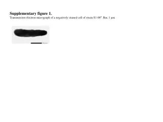
Supplementary figure 1. Transmission electron micrograph of a negatively stained cell of strain S1-08T. Bar, 1 μm.
Supplementary figure 2. Thin-layer chromatograms showing the total polar lipid compositions of (a) S1-05 and (b) S1-08T. Total polar lipids were detected by spraying the plate with 5 % molybdophosphoric acid. AL, unidentified aminolipid; GL, unidentified glycolipid; PE, phosphatidylethanolamine; L1, unidentified polar lipids. a) S1-05 L1 b) S1-08T GL1 GL2 S2 AL1 L1 PE AL2 GL1 S1 S2 GL2 AL1 PE AL2 S1