1 / 1

3C pro wt CM IEALFQ↓GPPKFR 3C pro mut CM IE K LFQ P PPKFR

DsRed2. GFP 2. 3C pro wt CM IEALFQ↓GPPKFR 3C pro mut CM IE K LFQ P PPKFR.

Télécharger la présentation

3C pro wt CM IEALFQ↓GPPKFR 3C pro mut CM IE K LFQ P PPKFR

An Image/Link below is provided (as is) to download presentation Download Policy: Content on the Website is provided to you AS IS for your information and personal use and may not be sold / licensed / shared on other websites without getting consent from its author. Content is provided to you AS IS for your information and personal use only. Download presentation by click this link. While downloading, if for some reason you are not able to download a presentation, the publisher may have deleted the file from their server. During download, if you can't get a presentation, the file might be deleted by the publisher.

E N D

Presentation Transcript


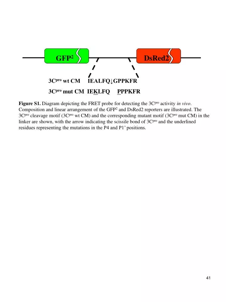
  1. DsRed2 GFP2 3Cpro wt CM IEALFQ↓GPPKFR 3Cpro mut CM IEKLFQ PPPKFR Figure S1. Diagram depicting the FRET probe for detecting the 3Cpro activity in vivo. Composition and linear arrangement of the GFP2 and DsRed2 reporters are illustrated. The 3Cpro cleavage motif (3Cpro wt CM) and the corresponding mutant motif (3Cpro mut CM) in the linker are shown, with the arrow indicating the scissile bond of 3Cpro and the underlined residues representing the mutations in the P4 and P1’ positions. 41

More Related