Download
1 / 9
90 likes | 204 Vues
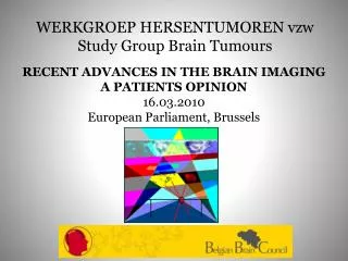
WERKGROEP HERSENTUMOREN vzw Study Group Brain Tumours. RECENT ADVANCES IN THE BRAIN IMAGING A PATIENTS OPINION 16.03.2010 European Parliament, Brussels. WERKGROEP HERSENTUMOREN VZW Study Group Brain Tumours. Brain Tumours Incidence HG BT 8 new cases/100.000 people/year

E N D
WERKGROEP HERSENTUMOREN vzwStudy Group Brain Tumours RECENT ADVANCES IN THE BRAIN IMAGING A PATIENTS OPINION 16.03.2010 European Parliament, Brussels
WERKGROEP HERSENTUMOREN VZWStudy Group Brain Tumours • Brain Tumours • Incidence HG BT 8 new cases/100.000 people/year • Incidence LG BT 8 new cases/100.000 people/year • Source: Improving Outcomes for People with Brain and other CNS Tumours; National institute for Health and Clinical Excellence, June 2006 In Europe 80.000 new cases/year
Typical Stages of the Disease ACUTE PHASE TREATMENT REHABILITATION Vague Symptoms Heavy Neurological Symptoms Diagnosis Detailed Diagnosis Neurosurgery Radiotherapy if HG Chemotherapy if HG Rehabilitation Adaptation-Integration Follow Up
LOOKING INSIDE THE SKULL MRI-SCAN -Work with magnetical resonance -Raise of temperature of 0,1 ° C -No known lasting effects -No evidence for cancer -Higher resolution • CT-SCAN • Work with X-rays • Ionising effects • May cause cancer (certainty) • Lower resolution 2 METHODS
DETAILED DIAGNOSIS • Primary or secundary brain tumour ? • Exact location • Functional analysis (PET-SCAN) • EEG • Stereotactical biopsy
NEUROSURGERY Stereotactical frame Mapping Awake surgery Neurostimulation Interventional MRI
FOLLOW UP Regular Controls with MRI
CONCLUSIONS Diagnosis, treatment and follow up depend entirely on the MRI technology MRI is a less harmful tool than CT Quality of images are superior with MRI