Download
1 / 111
1.2k likes | 1.63k Vues
DR.VINOD.G.V. HYPERTROPHIC CARDIOMYOPATHY. DEFINITION.

E N D
DR.VINOD.G.V HYPERTROPHIC CARDIOMYOPATHY
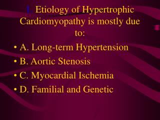
DEFINITION HCM is a disease state characterized by unexplained LV hypertrophy associated with nondilated ventricular chambers in the absence of another cardiac or systemic disease that itself would be capable of producing the magnitude of hypertrophy evident in given patient.
HCM is a common genetic cardiovascular disease Prevalance estimated to be 1:500
GENETICS • Caused by autosomal dominant mutations in genesencodingprotein components of the sarcomere and its constituent myofilament elements. • 1400 mutations identified among at least 8 genes.
Beta MHC mutations-clinical presentation apparent by late adolescents and develop substantial hypertrophy and more severe diseases. • MyBPC mutations can have delayed clinical presentation until age 50 or older.Less severe symptoms. • One Essential Myosin light chain mutation associated with mid cavitary hypertrophy.
cTnT mutations-modest hypertrophy, increased risk of sudden death • cTnI mutations- Greater predisposition of apical hypertrophy • Alpha tropomyosin-relatively good survival. Variable degree of hypertrophy
Insights From Genotype/Phenotype Studies • autosomal dominant disease -affects males and females equally. • Only 50% of the offspring of affected individuals will be at risk of inheriting the gene and developing the disease. • The offspring of unaffected family members carry no risk of inheriting the gene and developing the disease. • In any one family with FHCM, all affected members have the same mutation.
The onset of clinical manifestations is usually delayed until adolescence or early adulthood. • Clinical features of the phenotype are not predictive of sudden death, in certain genes there is a high correlation between the extent of ventricular hypertrophy and the incidence of sudden death. • Certain mutations are highly predictive of sudden death
MORPHOLOGY Asymmetric hypertrophy with small left ventricular cavity • Diffuse hypertrophy of septum and anterolateral free wall(70-75%) • Basal septal hypertrophy(10-15%) • Concentric hypertrophy(5%) • Apical hypertrophy(<5%) • Hypertrophy of lateral wall(1-2%)
MITRAL VALVE APPARATUS • twice the normal size due to elongation of both leaflets or segmental enlargement of only anterior leaflet or mid portion of posteror leaflet • Congenital and anomalous anterolateral papillary muscle insertion into the anterior leaflet without interposition of chordaetendineae and produce muscular midcavity outflow obstruction
Bizarre arrangement of muscle fibre bundles • Myocardial disarray consists short runs of severly hypertrophied fibres interrupted by connective tissue • Myocardial fibrosis with degenerating muscle fibres • “Whorling” of muscle fibres • Volume of interstitial collagen increases
Abnormal intramural coronary arteries with thickened wall and narrow lumen near to areas of replacement fibrosis • Microvascular disease -silent myocardial ischemia - myocyte death replacement fibrosis often transmural
PATHOPHYSIOLOGY LVOT OBSTRUCTION • Produced by SAM of mitral valve and midsystolic ventricular septal contact • SAM is abrupt anterior motion of the mitral valve in which elongated leaflets move toward the septum with a sharp-angled 90 degree bend and generated largely by a drag effect, ie hydrodynamic pushing force of flow directly on the leaflets
In the classic form of obstructive HCM, the obstruction will occur at the most basal portion of the septum as it projects into the left ventricular outflow tract. the obstruction may also extend into the left ventricle from SAM of the chordal apparatus. patients with midventricular obstruction hypertrophied papillary muscle abuts against the ventricular septum.
Most patients will have SAM of the anterior leaflet, but this may also occur with the posterior leaflet. • The exact site of the obstruction may be determined by visualizing the region of the SAM-septal contact
Obstruction is dynamic-varying with loading conditions and contractility of LV • Increase in contractility VPC Dobutamine,Isoproterenol Exercise • Decrease in afterload/volume Valsalva maneuver Nitroglycerine/amylnitrite inhalation Blood loss dehydration
Definitions of Dynamic Left Ventricular Outflow Tract Obstruction
DIASTOLIC DYSFUNCTION • Impaired relaxation,filling and increased ventricular stiffness • Contributing to the symptoms • Rapid filling phase is prolonged • Decreased rate and volume of filling • Compensatory increase in atrial filling • Decrease compliance is due to hypertrophy,replacement scarring ,interstitial fibrosis and disorganised cellular architecture
MICROVASCULAR DYSFUNCTION MYOCARDIAL ISCHEMIA • Myocardial ischemia is unrelated to epicardial coronary artery disease. supply demand mismatch due to hypertrophy. abnormally small and partially obliterated intramural coronary arteries • Ischemia causes myocardial scaring and remodelling and replacement fibrosis which is a determinent of progressive heart failure and substrate for arrythmia
MITRAL REGURGITATION • Common in patients with LVOT obstruction. • Secondary to distortion of mitral valve apparatus from SAM. • The jet of MR is directed laterally and posteriorly and predominantly during late and mid systole • Severity proportional to LVOT obstruction.
CLINICAL FEATURES Symptoms • Majority are asymptomatic • Dyspnoea occurs 90% of symptomatic patients • Syncope and presyncope in 20 and 50% respectively due to either hemodynamic or rhythm abnormality.
Angina -70-80% small artery narrowing intramural compression of small arteries from myocardial hypertrophy abnormal diastolic filling oxygen supply demand mismatch abnormal coronary flow reserve
PHYSICAL EXAMINATION Classic findings applied to patients with LVOT Obstruction • carotid pulse is brisk with spike and dome pattern with a rapid rise (percussion wave) followed by a mid systolic drop inturn followed by a secondary wave(Tidal wave) • Apical impulse:double or triple
Second heart sound:paradoxical split 20% • Fourth heart sound is present • Murmer: cresendo-decresendo at left sternalborder.radiates to base as well as apex. Seldom radiates to carotid arteries Dynamic auscultation: maneuvers that decrease preload will increase the dynamic gradient and increase intensity of murmer. Eg:standing and strain phase of valsalva
ECG • Abnormal in 95% of HCM patients • LVH seen in 70-80% patients • Abnormal Q waves simulating myocardial infarction due to disturbance of activation of ventricular septum • Apical HCM:Diffuse symmetric T wave inversion across precordium • Atrial fibrillation:25-30% of older patients
CARDIAC CATHETERIZATION "pull-back" pressure tracing systolic gradient between the apex and base. • the small left ventricular cavity hyperdynamic systolic function • catheter "entrapment" may occur resulting in a falsely increased left ventricular systolic pressure
ideally assessed by a simultaneous left ventricular inflow and left ventricular outflow (or aortic) pressure. • inflow position avoids the problem of catheter entrapment and is best obtained by a transseptal approach.
BROCKENBROUGH PHENOMENON useful for latent obstruction • After a premature contraction- increase in the contractility of the ventricle marked increase in the degree of dynamic obstruction.
increase in gradient and a decrease in the aortic pulse pressure after the pause. • fixed obstruction - increase in gradient from the increase in stroke volume increase in aortic pulse pressure
LEFT VENTRICULOGRAPHY • small left ventricular cavity size with hypertrophied papillary muscles • Hyperdynamic systolic function - complete obliteration of the mid and apical cavity in systole • apical HCM - fixed obliteration of the apex by the hypertrophied muscle- "spade-like" configuration.
midventricular obstruction an apical akineticdyskinetic pouch with"aneurysm" formation
STRESS TESTING Important adverse prognostic factors decrease in blood pressure appearance of ventricular arrhythmias. • Exercise is the most physiologic form of provocation to attempt to detect latent LVOT obstruction.
CMR More accurate than echo Can detect 6% more hypertrophy Accurate measurement of thickness Should be done in Poor echo window Discrepancy between Clinical findings / ECG / Echo
CMR - Poor Prognostic factors • Markedly elevated LV mass index (men > 91 g/m2, women > 69 g/m2) - sensitive(100%) • Maximal wall thickness of more than 30 mm specific (91%) for cardiac deaths
Type 1:Anterior segment of septum(10%) • Type 2:Both anterior and posterior segment(20%) • Type 3:Septum and anterolateral free wall(52%) • Type 4:Other regions including apical HCM (18%)
Right ventricular (RV) hypertrophy • Myocardial edema by T2-weighted imaging • LGE has been associated with • Ventricular arrhythmias • Progressive ventricular dilation
BURNTOUT HCM • 3% manifest the end stage- systolic dysfunction (ejection fraction <50%) • Progressive heart failure • Often associated with AF • Patterns of LV remodeling • Wall thinning and cavity dilation,