Download
1 / 65
650 likes | 673 Vues
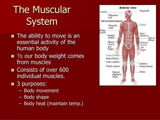
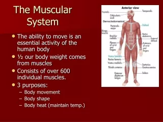
The Muscular System. Biol 105 Lecture 12 Chapter 6. Outline. Characteristics of muscles Three types of muscles Functions of muscles Structure of skeletal muscles Mechanics of muscle contraction Energy source for muscle contraction. Muscular System.

E N D
The Muscular System Biol 105 Lecture 12 Chapter 6
Outline • Characteristics of muscles • Three types of muscles • Functions of muscles • Structure of skeletal muscles • Mechanics of muscle contraction • Energy source for muscle contraction
Muscular System • Remember there were different types of muscles: cardiac, smooth and skeletal. • All muscle cells are elongated and therefore are called muscle fibers. • All muscle tissues contract. • Muscles contain muscle cells (called muscle fibers), connective tissue, blood vessels, and nerves
Types of Muscles • Smooth muscle • Cardiac muscle • Skeletal muscle 11-2
Smooth muscle • Smooth muscles are involuntary muscles found in the walls of many internal organs (digestive tract, respiratory system, blood vessels). • Function to aid in the function of other organs 11-2
Cardiac muscle • Cardiac muscles are involuntary muscles found only in the heart wall. • Functions by contracting to force blood from the heart into the arteries 11-2
Skeletal muscle • Skeletal muscleare voluntary muscles attached to the skeleton. • Usually work in pairs 11-2
Skeletal Muscles Work in Pairs • Most skeletal muscles are antangonistic pairs. • One muscle contracts, the other relaxes • Muscles are attached to the bone by tendons • Skeletal Muscles are usually attached to two bones on opposite sides of a joint
Skeletal Muscles Work in Pairs • The origin of the muscle is attached to the bone that remains stationary during movement • The insertion is attached to the bone that moves • Bones act as levers in working with skeletal muscles to produce movement
Skeletal Muscles Work in Pairs Origin of muscle: attachment of muscle to less moveable bone The biceps contracts and pulls the forearm up, flexing the arm. The relaxed triceps is stretched. Insertion of muscle: attachment of muscle to more moveable bone (a) Flexion Figure 6.1a
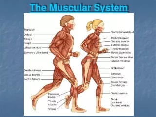
Functions of Skeletal Muscles • Support the body – maintain our posture • Movement of bones, and other tissues • Help maintain a constant body temperature – generates heat • Helps move blood through the veins and lymphatic fluid through the lymphatic vessels • Help to protect vital organs and stabilize joints
Smooth muscles are under this kind of control • Voluntary • Involuntary
Smooth muscles are found in • The heart • Digestive tract • Attached to bones
Structure of Skeletal Muscles • Muscles are covered by connective tissue called fascia. • A muscle contains bundles of skeletal muscle fibers (muscle cells), the bundles are called fascicles. These bundles are covered by connective tissue. • Blood vessels and nerves are between the fascicles.
Structure of Skeletal Muscles Skeletal muscle consists of many bundles of muscle cells. A muscle cell consists of many myofibrils. A bundle of muscle cells is called a fascicle. (a) A section of a skeletal muscle The striped (striated) appearance of a skeletal muscle cell is due to the regular arrangement of myofilaments. (b) A light micrograph of a longitudinal view of skeletal muscle cells Figure 6.3a–b
Sarcomeres The striped (striated) appearance of a skeletal muscle cell is due to the regular arrangement of myofilaments. (b) A light micrograph of a longitudinal view of skeletal muscle cells Z line One sarcomere (c) A diagram and electron micrograph of a myofibril Figure 6.3b–c
A bundle of muscle cells is called: • Fascicles • Fascia • Muscle Fibers
Muscle Cells • Muscle cells are long cells called muscle fibers. • The muscle fiber is composed of long thin myofibrils
c. myofibril a. T tubule b. Sarcoplasmic reticulum d. Z line e. sarcomere f. sarcolemma
Muscle Cells cont • Myofibrils are bundles of myofilaments that contracts. • Myofilaments are made of actin and myosin filaments. • When muscle fibers are stimulated to contract, myofilaments slide past one another, causing sarcomeres to shorten.
Muscle Cell Components • Muscle cells (muscle fibers) have many of the same components as typical cells have but some of their components have different names
Muscle Cell Components • Sarcolemma – plasma membrane (cell membrane) • Sarcoplasm – similar to cytoplasm, contains large amount of stored glycogen and myoglobin. • Myoglobin is an oxygen binding protein similar to hemoglobin, but found only in muscles • Sarcoplasmic reticulum – similar to endoplasmic reticulum, one of its functions is to store Ca2+
Muscle Cell Components • Muscle cells (muscle fibers) also have unique features: • Multiple nuclei • Transverse tubules (T tubules) – extensions of the sarcolemma that come into contact with the sarcoplasmic reticulum.
Muscle Contraction • The small myofibrils that make up the muscle fiber (muscle cell) contain two types of myofilaments: actin and myosin filaments • Sarcomere is the name for the structural unit of these myofilaments • The sarcomere goes between two dark lines = Z lines. The Z lines are protein sheets where the actin filaments attach
Sarcomeres Z line One sarcomere (c) A diagram and electron micrograph of a myofibril Z line Z line One sarcomere Actin Myosin (d) A sarcomere, the contractile unit of a skeletal muscle, contains actin and myosin myofilaments. Figure 6.3c–d
Myofilaments – actin and myosin • The two myofilaments are: • Actin filaments: thin filaments that formed by two intertwining strands of the protein actin. • Myosin filaments: Thick filaments of the protein myosin shaped like a golf club, with a round “head”.
Myofilaments – actin and myosin • The myosin heads can bind and detach from the thin actin filament. When bound it creates cross-bridges. • When the muscle is stimulated, these filaments slide past each other, making the sarcomere to shorten
Muscle Contraction cont • A neuron signals the muscle to contract • The myosin heads attach to the actin then pull the actin toward the center of the sarcomere • Then the myosin heads detach
Sarcomeres Figure 6.4
Neuromuscular Junction Figure 6.7 (1 of 2)
Steps of Muscle Contraction • Action potentials are transmitted through the neurons. • At the end of the neurons neurotransmitters are released • Neurotransmitters bind to receptor on the sarcolemma
Steps of Muscle Contraction • The receptors are ion channels that open • An action potential travels through the T tubules • The action potential goes to the sarcoplasmic reticulum • The sarcoplasmic reticulum releases Ca2+.
Steps of Muscle Contraction • The calcium binds to the troponin on the actin filament • This opens up binding site for the myosin to attach • Now the myosin binds to the actin • ATP is needed for the myosin to slide past the actin
Sarcomeres Figure 6.6 (1 of 2)
Sarcomeres Figure 6.6 (2 of 2)
Tropomyosin-troponin complex • The tropomyosin-troponin complex is attached to the actin filament. • Calcium binds to the troponin, causing a shift in the complex, opening the sites for myosin to attach.
What is the an oxygen binding protein found only in muscles? • Myosin • Actin • Hemoglobin • Myoglobin
What ion is required for the myofilaments to bind to each other? • Potassium • Calcium • Chloride • Sodium
Where is the calcium stored? • Nucleus • Sarcolemma • Sarcoplasmic reticulum
c. myofibril a. T tubule b. Sarcoplasmic reticulum d. Z line e. sarcomere f. sarcolemma
ATP • ATP is the currency. Like money in the bank. • The bonds between the phosphate groups are high energy bonds