Download

1 / 71
720 likes | 1.07k Vues
Foot, Ankle, Lower leg. Lesson 1 – bone and ligament anatomy. Foot bones. Foot bones 26 bones 14 phalangeal 5 metatarsals 7 tarsals. Toes. Each toe except for the big toe has three phalanges Toes 2-5 are also known as digits The big toe, a.k.a. hallux, has two.

E N D
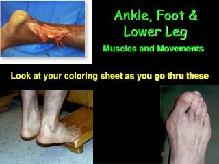
Foot, Ankle, Lower leg Lesson 1 – bone and ligament anatomy
Foot bones • Foot bones • 26 bones • 14 phalangeal • 5 metatarsals • 7 tarsals
Toes • Each toe except for the big toe has three phalanges • Toes 2-5 are also known as digits • The big toe, a.k.a. hallux, has two. • The toes are designed to widen our base for balance and propelling our body. • Two sesamoid bones are located under the 1st metatarsalphalangeal joint (MTP joint) • These bones help increase the mechanical advantage of the flexor tendons that run under the big toe.
Metatarsals • Five bones that lie between the toes and the tarsal bones • The first metatarsal is the biggest and the strongest. • This helps it function as the main weight bearing support during walking and running. • The fifth metatarsal protrudes on the lateral aspect of the foot. • Place where ligaments and muscle tendons attach
Tarsal bones • Calcaneus • Largest of tarsal bones • Shapes the heel and sits below the talus • Conveys body weight to the ground • Attachment for achilles tendon and several structures on plantar side • Palpate on the posterior bottom of the foot
Tarsal bone cont… • Talus • Located above the calcaneus • Fits between the lateral and medial malleoli forming the talocural joint (a.k.a ankle joint) - mortise • Irregular shaped • The bone is broader anterior than posterior • Which gives the ankle more stability in dorsiflexion as it makes a tight fit between the malleoli (10 deg in DF vs. 23 deg in PF)
Tarsal bones cont. • Navicular • Located anterior of the talus on the medial aspect of the foot. • Small tubercle may be palpated on the medial foot. • Anteriorly articulates with the cuneiforms • Cuboid • Located on lateral aspect of foot. (palpate on lateral foot) • Posterior articulates with the calcaneus and anterior with the 4th and 5th metatarsals • Cuneiforms • Three bones located between the navicular and 3-5 metatarsals • Palpate from mid foot to the medial side
Lower leg bones • Tibia • Second longest bone in body • Principle weight bearing • Located on medial side of lower leg • The shaft has three sides – posterior, medial, lateral • Lateral and posterior covered by muscle • Triangle shaped on the top and round near the bottom • Causes a anatomical weakness – bone more dense in this area • Forms medial malleolus
Lower leg bones cont. • Fibula • Long slender bone located on lateral leg • Non weight bearing • Primary function is to provide attachment for muscles • Distal portion forms the lateral malleolus • Malleolus extends further distally than medial to give it more stability
Arches of the foot • Medial longitudinal arch • Runs along the medal side of the foot from the calcaneus to distal head of the first metatarsal • Bony support are the medial bones • The soft tissue support is the plantar calcaneolnavicular ligament (a.k.a spring ligament) and posterior tibialis muscle • Lateral Longitudinal arch • Runs along lateral portion of the foot. • Much lower and less flexible than the medial
Arches cont… • Transverse arch • Half dome over the anterior portion foot over the tarsal bones, primarily the cuboid and internal cuneiform • Anterior metatarsal arch • Shaped by the distal heads of the metatarsal heads
Foot ligaments • Spring ligament • a.k.a plantar calcaneonavicular ligament • Located on medial side of foot • Gives support to the medial arch • Helps with shock absorption • Bifurcate ligament • Located on lateral side of foot • Located under fat pad on foot • Connects cuboid to calcaneus and calcaneus to the navicular
Plantar fascia • Lies on the bottom of the foot from the calcaneus to the head of each metatarsal head • Helps support the foot against downward forces
Ankle/lower leg ligaments • Interroseous membrane • Connective tissue that connects the tibia and fibula • The membrane fills the whole space between the tibia and fibula in the lower leg • The membrane helps diffuse forces placed on the lower leg
Lateral ankle ligaments • Anterior/Posterior talofibular ligaments • Both are located on lateral ankle • Helps prevent against anterior/posterior torsion and inversion of the ankle (talus specifically) • Calcaneofibular ligament • Located laterally it traverses inferiorly of the lateral malleolus • Protects against inversion of the calcaneus
Lateral ankle ligaments cont… • Calcaneofibular ligament • Located laterally it traverses inferiorly of the lateral malleolus • Protects against inversion of the calcaneus
Ankle/lower leg ligaments • Anterior/posterior tibiofibular ligaments • Sometimes called the syndesmotic ligaments • They connect the tibia and fibula together at the distal end of the bones – forms the distal portion of the interroseous membrane
Medial ankle ligaments • Deltoid ligaments • Located medially on the ankle • Technically three ligaments – treat as one • Triangle shape that begins on the medial malleolus and ends on the medial talus, calcaneus, and navicular bone • Protects against eversion, pronation, and anterior displacement of the ankle (talus specifically)
Foot, ankle, and lower leg Lesson 2 – muscles, movement and other structures
Lateral muscles • Peroneal brevis • Origin or proximal attachment - lower 2/3 of outer surface of fibula • Insertion or distal attachment – base of 5th metatarsal • Action – eversion of foot and plantar flexion • Peroneal Longus • Origin – upper 2/3 of fibula • Insertion – undersurface of medial cuneiform and 1st metatarsal • Action – plantar flexion and eversion
Medial muscles • Tibialis posterior • Proximal attachment – posterior surface of tibia, fibula, and interroseous membrane • Distal attachment – undersurface of navicular, cuneiforms, and base of 2-4 metatarsals • Action– inversion and plantar flexion of foot/ankle • Flexor Hallicus Longus • Proximal attachment – Lower 2/3 of posterior fibula • Distal attachment – Undersurface of base of distal phalanx of the Big toe (1st) • Action – Plantar flexion of big toe and inversion and plantar flexion of foot/ankle • Flexor Digitorum longus • Proximal attachment – Lower 2/3 of posterior tibia • Distal attachment – Base of distal phalanx of toes 2-5 • Action – plantar flexion of toes 2-5 and plantar flexion and inversion of ankle/foot
Anterior Muscles • Anterior Tibialis • Proximal attachment – Upper 2/3 of anterior tibia • Distal attachment – inner surface medial cuneiform and 1st metatarsal • Action – Dorsal flexion of ankle and inversion of foot • Extensor Hallicus longus • Proximal attachment – Anterior/inner surface of middle 2/3 of fibula • Distal attachment – top of distal phalanx of big toe • Action – dorsal flexion of ankle and big toe and foot inversion • Extensor Digitorum longus • Proximal attachment – Lateral condyle of Tibia, head of fibula, and upper 2/3 of fibula • Distal attachment – top of middle and distal phalanx of toes 2-5 • Action – Dorsal flexion of ankle and toes 2-5, eversion of foot.
Posterior Muscles • Gastrocnemius • Proximal attachment – posterior surface of medial and lateral condyles of femur • Distal attachment – posterior surface of the calcaneus • Action – plantar flexion of the ankle and flexion of the knee • Soleus • Proximal attachment – Upper 2/3 of posterior surface of tibia and fibula • Distal attachment – posterior surface of calcaneus • Action – plantar flexion of the ankle • Achilles tendon – formed from the gastrocnemius and soleus
Other structures • Anterior and Posterior tibial arteries • Main blood supply for the ankle and foot • Located in the anterior portion of the foot and behind medial malleolus • Common peroneal nerve • Located on lateral side • Tibial nerve • Located behind medial malleolus • Retrocalcaneal bursa • Located under the achilles tendon attachment • Retinaculum • Fascia that holds down ankle tendons as they curl from the lower leg into the foot keeping them in place • Joint capsule • Fascia tissue that encompasses the ankle
Foot, ankle, and Lower leg Lesson 3 – foot injuries
Heel Bruise • Contusion to the calcaneus • Etiology • Landing directly on the heel without or with limited protection • Signs and symptoms • Pain, pt tenderness, and swelling over calcaneus • Difficulty walking • Management • RICE immediately • Use of crutches for first few days • Use of donut pad when beginning walking • Use of other modalities; US, whirlpool, ect for help in healing.
Base of Fifth Tendonitis • Inflammation of Peroneal brevis at the insertion point at the base of the fifth metatarsal. • Etiology • Overuse from running, poor support in shoes, complication of ankle sprain. • Signs and symptoms • Pain, point tenderness, and swelling over base of fifth. • Difficulty walking • Weakness and pain with eversion • Management • RICE • Other modalities – e-stim, ultrasound, WP • Walking boot if becomes to severe • Complications • If not treated and cured quickly can lead to avulsion of Peroneal tendon off base of fifth (Jones fracture) – leads to surgery
Retrocalcaneal Bursitis • Bursa irritation over the Achilles/calcaneal insertion • Etiology • Overuse – running, jumping • Symptoms • Pain and swelling on insertion • Strength loss minimum • Pain with passive dorsiflexion • Treatment • RICE – similar too Severs disease
Sesmoiditis • Irritation to sesmoid bones under 1st metatarsal/phalangeal joint • Etiology • Running or jumping – excessive force on joint • Symptoms • Pain and swelling over joint • Pain with passive extension • Pain with active flexion • Treatment • Bracing – tape - donut • Ice and other modalities
Plantarfascitis • Irritation of fascia that lays on the plantar foot – especially around ½” from base of calcaneus • Etiology • Overuse – arch taking to much pressure. Not enough padding on heel of shoes • Running on toes instead of heel to toe • Poor support of arch • Symptoms • Pain around calcaneus ½” from origin. Hurts immensely first thing in the morning. • Treatment • Arch taping • Getting proper shoes • Ice massage • Stretching • Takes along time to heal
Turf Toe • Hyperextension or hyperflexion of 1st metatarsal • Etiology • Forceful flex or over extension of big toe while running • Stubbing toe • Symptoms • Swelling and pain over joint • Treatment • RICE • Tape • Modalities
Bifurcate sprain • Sprain of ligament that holds talus, cuboid and navicular together • Located around fat pad on lateral side of foot • Etiology • Inversion of foot • symptoms • similar to ankle • swelling and pain over fat pad in foot and lateral foot • pain and mild weakness with eversion • Treatment • Similar to ankle • RICE • Foot strengthening • Heals faster than regular ankle sprain
Medial and Lateral arch • Etiology • Poor shoes, overweight, postural anomalies, weakened support structures • Signs and symptoms • Soreness, tiredness around arch • Pain when running • Management • Tape • Strengthening of surrounding structures
Fallen Metatarsal heads • Etiology • Weakened surrounding structures due to undue stress being placed on toes 2-5 causing them to splay apart. • Signs and symptoms • Transverse arch becomes flattened and may see one or two head of metatarsals depress • Pain when walking • Management • Place small pad behind fallen metatarsal head helping push back up • Strengthen surrounding structures
Fractures • Dome of Talus • etiology • severe inversion or forceful dorsiflexion • signs and symptoms • pain located in center of ankle in mortise area • management • RICE • refer to doctor
Fractures • Navicular • Etiology • high arches • poor shoes • signs and symptoms • pain when running and jumping around Navicular • point tender over tubercle • management • RICE • x-rays • walking boot