Optic Disc Evaluation IN Glaucoma
Optic Disc Evaluation IN Glaucoma. Dr Deepak Megur DOMS FRCS Ed Glaucoma & Cataract Services Megur Eye Care Centre Bidar 585401. What is Glaucoma…?. Glaucoma =Optic neuropathy. The evaluation of the appearance of the optic disc is central to the diagnosis and management of Glaucoma


Optic Disc Evaluation IN Glaucoma
E N D
Presentation Transcript
Optic Disc Evaluation IN Glaucoma Dr Deepak MegurDOMS FRCS Ed Glaucoma & Cataract Services Megur Eye Care Centre Bidar 585401
Glaucoma =Optic neuropathy • The evaluation of the appearance of the optic disc is central to the diagnosis and management of Glaucoma • Optic Disc Evaluation: • Why..? • How..? • What to look for..?
The 4 goals of optic disc evaluation • Distinguishing between the healthy and the sick =Diagnosing. • Quantifying the amount of damage: Healthy,Mild,Moderate,Advanced Disease • MonitoringChange,for better or for worse • Quantitating the rate of change
3 mm 1.5 mm
Optic Disc Evaluation .. • Slit lamp biomicroscopy : Ideal • Stereoscopic View –Cupping • Measuring the optic disc size • Direct Ophthalmoscopy • Good Magnification • Indirect Ophthalmoscopy • Overall View • Optic disc Photoghraphy. • Documentation,Monitoring for progression.
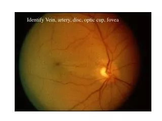
The 7 parameters to look for… • Disc:Size and Shape • 2)Neuroretinal Rim (NRR): • Size,Shape,Pallor • ISNT rule • 3)Cup: Size and Shape in relation to the optic disc size, • -Vertical C/ D Ratio, Cup depth / Excavation • 4)Optic DiscHemorrhage: Presence & Location
The 7 parameters to look for… 5) Nerve Fibre Layer Defect: • focal & diffuse 6) Para Papillary Atrophy; • Size,location & Configuration 7) Retinal Arterial Attenuation: • focal & diffuse
All these variables can be measured semiquantitavively by ophthalmoscopy without applying sophisticated techniques
1)Optic Disc: Size & Shape • Determining the size of the disc =Crucial • Helps to differentiate Physiological cupping from Pathological. • Large discs have big physiological cups. • Small Discs have small cups or no cups • Measurement of Vertical Disc diameter : • Length of the vertical beam of slit lamp light • Multiplied by correction factor of the condensing lens • Volk 60 D= X 1 • Volk 90D= X 1.5
Cup: Size, Shape, location in relation to the disc size • Optic Cup= Excavation in the optic nerve head • Stereoscopic evaluation • In normal eyes= Areas of optic disc & Optic cup are corelated • Large optic discs=Large cup • Small optic disc =Small cup or no cup • Early & moderate glaucomatous damage in small disc may be missed because of the erroneously low cup disc ratios
3 mm 1.5 mm
Early & moderate glaucomatous damage in small disc may be missed because of the erroneously low cup disc ratios
Vertical Cup Disc Ratio • Vertically oval optic disc • Horizontally oval optic cup • In normal eyes: Horizontal CD ratio > than vertical CD ratio • In Glaucomatous eyes: Vertical CD ratio > than the horizontal CD ratio
The Neuroretinal Rim • Size, Shape, Pallor. • The ISNT rule: I>S>N>T
Thinning of the NRR • Pallor of NRR • Notching: • A notch is a localized defect in the Neuroretinal rim on the cup side of the rim
The Neurretinal rim loss in Glaucoma • Usual sequence of NRR loss in Glaucoma: • Inferotemporal • Superotemporal • Horizontal temporal • Inferonasal • Superonasal • In contrast,in the non glaucomatous optic nerve damage, the NRR is not always affected and hence contour of NRR is maintained.
NRR , the “ISNT Rule” I>S>N>T I>S>N>T
I<S>N>T I<S<N>T
I<=S>n>T I>S>N>T
Splinter or Flame shaped hemorrhages At the margin of the disc Hallmark of Glaucomatous optic nerve damage 4 to 7 % of eyes with galucoma Found in early & moderately advanced Glaucoma and rare in very advanced stage Located usually in the inferotemporal & superotemporal disc margins Associated with localized RNFL defect and neuroretinal rim notches . SuggestsProgression. More common in NTG Optic Disc Hemorrhage
Retinal Nerve Fibre Layer Defect • RNFL contains retinal ganglion cells axons covered by astrocytes and bundled by processes of muller cells • Seen as bright fine striations fanning off from the disc to the periphery. • Dilated pupil, green light, clear optical media aids the evaluation of RNFL
Retinal Nerve Fibre Layer Defect • Localized RNFL defects: • Can be detected before visual field defect has developed • Focal type of NTG • Early to medium advanced Glaucomatous damage • Diffuse loss of RNFL: • More difficult to detect • Peripapillary retinal vessels appear bare • Underlying Choroidal vessels more clearly seen .
Parapapillary Chorioretinal atrophy • 2 zones • Central Beta zone • Peripheral alpha zone • Beta zone occurs more often in glaucomatous eyes than in normal eyes. • Helps to differentiate various subtypes of POAG • Helps to differentiate from nonglaucomatous optic nerve damage
Diffuse narrowing: Decreasing NRR Increased RNFL loss Increased Visual field defects Focal Attenuation More common in NTG Degree of narrowing increases with amount of damage. Retinal Artery attenuation
Pre Perimetric Diagnosis of Glaucomatous Optic Nerve damage • Most important Variables • Shape of the NRR • Size of the cup in relation to the optic disc • Diffuse or focal RNFL defects • Disc Hemorrhages
Moderate One pole of the disc is damaged
Advanced Both the poles affected
Optic Disc PhotographsOptic Disc Drawings Documentation of disc damage: • Monitoring change for progression • Rate of change