Download
1 / 90
900 likes | 1.01k Vues
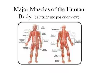
Advanced Biology Ch 10:Muscle Tissue Ch 11: The Muscular System. 3 types: 1) skeletal -pulls on bones to cause movement 2) cardiac -pumps blood thru circulatory system 3) smooth -pushes fluids & solids thru body, regulates blood vessel diameter. Skeletal Muscle: Functions:

E N D
Advanced Biology Ch 10:Muscle Tissue Ch 11: The Muscular System
3 types: • 1) skeletal-pulls on bones to cause movement • 2) cardiac-pumps blood thru circulatory system • 3) smooth-pushes fluids & solids thru body, regulates blood vessel diameter
Skeletal Muscle: • Functions: • a) Produce skeletal movements • -contract & tendons pull on bones
b) Maintain posture & position • -constant tension keeps head in position & body over feet • c) Soft tissue support • -muscles lining abdomino- pelvic cavity support weight of organs & offer protection
d) Guard entrances & exits • -closes openings in digestive & urinary tracts • e) Produce heat • -contractions require energy that is converted into heat to maintain temp.
f) Nutrient reserve storage • -proteins in muscles are broken down to provide amino acids for enzymes & energy
Organization • -3 connective tissue layers • 1) Epimysium: • -surrounds entire muscle • -separates muscle from other tissues/organs
2) Perimysium: • -divides muscle into compartments • -each contains a fascicle- bundle of muscle fibers • -each receives a branch of blood vessels & nerves
3) Endomysium: • -surrounds individual muscle fibers (cells)
Collagen fibers from each layer join to form a tendon (connects muscle to bone) or aponeurosis (broad sheet that connects muscle to bone)
Microscopic structure • -cells can be 30 cm long & 100 µm (.1 mm)diameter • -very large compared to most cells • -multinucleate • -may be 100’s/cell • -lie just below membrane
-Sarcolemma-plasma membrane of muscle fiber • -Sarcoplasm-cytoplasm
-Transverse (T) tubules- narrow tubes, continuous w/ sarcolemma that extend thru sarcoplasm • -fluid-filled • -contain same electrical charge as sarcolemma • -send impulse to entire fiber so it all contract together
-Myofibrils- cylindrical structures that run the length of a fiber • -made of myofilaments: thin strands/filaments of protein • -2 types • 1)Thin filaments-actin • 2)Thick filaments-myosin
-can shorten & are responsible for muscle contracting • -anchored to ends of sarcolemma which eventually becomes a tendon which pulls on bone • -space filled w/ mitochondria & glycogen
-Sarcoplasmic reticulum- membrane complexs, similar to smooth ER • -surround each myofibril between T-tubules • -terminal cisternae- expanded, fused chambers where SR meets T-tubule
-triad-2 terminal cisternae & a T-tubule • -cells pump CA2+ ions out of cells, they also transport them into the terminal cisternae • -may have 1000x higher concentration of free Ca2+
-calsequestrin (protein) binds Ca2+ • -total Ca2+ may be 40,000x greater • -contractions begin when Ca2+ ions are released into sarcoplasm
-Sarcomere- repeating, individual contractile units in myofibrils • -1 myofibril can have 10,000 • -4 components:
1) Thick filaments-myosin w/ associated titin (elastic filaments) • 2) Think filaments-actin • 3) Stabilizing proteins • 4) Regulating proteins • -create banded/striated appearance
I-band-light band (only actin) • A-band-dark (myosin & actin) • H-zone- only myosin
M-line-proteins that stabilize position of thick bands • Zone of overlap-thin filaments between thick • -6 thin surround a thick • -3 thick surround a thin • p. 289
Z-line-boundary between sarcomeres • -proteins that connect thin filaments • -titin proteins extend from thick filaments & attach to Z-line • -helps filament alignment
-Surrounded by 2 T-tubules & triads located at zones of overlap
-Thin filaments: • -each contains 4 proteins • -active site: area where myosin can bind • -covered by troponin- tropomyosin complex when a muscle is relaxed
-in resting muscle, intracellular Ca2+ concentrations are very low & binding site is empty • -Troponin-tropomyosin complex must move in order for contraction to occur • -calcium triggers process
-Thick filaments • -contain 300 twisted myosin molecules • -tail-where molecules are bound to each other • -head-projects toward actin • -made of 2 globular protein subunits
-interact w/ active site to form cross-bridges • -hinge that allows head to pivot freely • -arranged so tails point toward M-line • -core contains titin that extends past fiber & attaches to Z-line
Contraction • Events: • 1)H-zones & I-bands get smaller • 2)Zones of Overlap get larger • 3)Z-lines get closer together • 4)A-band width stays constant
Sliding Filament Theory-thin filaments slide toward M-line moving across thick filaments, causing sarcomere to shorten & muscles to contract
Control of Muscle Activity • -nerves from central nervous system (brain & spinal cord) control contractions • -Neuromuscular junction: area where nerve connects/ communicates w/ muscle fiber • -1/fiber
-ends w/ synaptic terminal • -filled with acetylcholine (Ach)-a neurotransmitter that is released by the neuron from the synaptic terminal, attaches to muscle fiber & alters the permeability of the sarcolemma
-Synaptic cleft: space between synaptic terminal & muscle sarcolemma • -Motor end plate: sarcolemma opposite synaptic terminal that houses receptors where Ach binds • -folded to surface area
-Acetylcholinesterase: (AChE)-enzymes that breaks down ACh, found in synaptic cleft & sarcolemma
-Steps of Stimulation • 1) Action potential: (electrical impulse to signal release of ACh) reaches synaptic terminal • 2) ACh is released thru exocytosis into synaptic cleft
3) ACh binds to motor end plate receptors & changes permeability to Na+ ions, which move into sarcoplasm until AChE removes ACh from receptor sites
4) Na+ movement creates an action potential in sarcolemma, moves inward thru T-tubules • 5) ACh broken down by AChE & system is ready for another action potential
Excitation-Contraction Coupling: process that uses the stimulus from an action potential to create a muscle contraction • -occurs at triads
-action potential causes cisternae to release Ca2+ ions into sarcoplasm at a sarcomere’s zone of overlap • -normally troponin (enzyme) covers the active site of actin strands, Ca2+ causes troponin to release & allow for myosin heads to bond
Contraction cycle: • 1) Troponin removed & active sites available • 2) Myosin head binds to active site forming a cross-bridge (requires 1 ATP)
3) Myosin heads pivot from being pointed away from the M-line to being pointed towards it (power stroke) • 4) Cross-bridges detach (myosin head lets go of active site)
5) Another ATP is split to re- energize free myosin head which causes it to pivot back to its original position • -Cycle begins again as long as Ca2+ is present & ATP is available • -Each power stroke shortens the sarcomere by 1%
-Length of contraction depends on: • 1) Length of stimulation at neuromuscular junction • 2) Availability of free Ca2+ ions in sarcoplasm • 3) Availability of ATP
-ACh binding occurs only briefly, so action potentials must continue to be applied in rapid succession to maintain a contraction • -Once action potential ends, sarcoplasmic reticulum permeability changes & absorbs Ca2+, active site is recovered
-at death, no more nutrients/ oxygen cycles, Ca2+ enters sarcoplasm which triggers contraction. Cross-bridges can’t detach because there’s no ATP-causes rigor mortis-constant contraction of skeletal muscles at death, lasts 15-25 hours until autolytic enzymes break down Z-lines & titin