Download
1 / 46
550 likes | 1.02k Vues
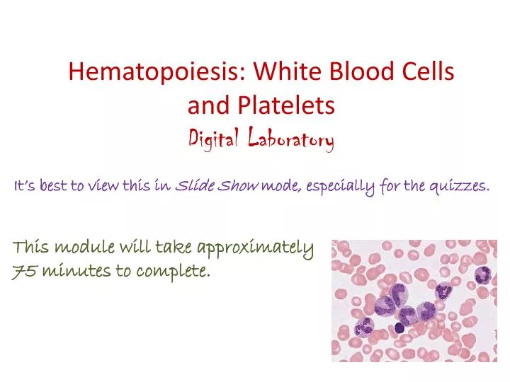
Hematopoiesis: White Blood Cells and Platelets Digital Laboratory. It’s best to view this in Slide Show mode, especially for the quizzes. This module will take approximately 75 minutes to complete. After completing this exercise, you should be able to:

E N D
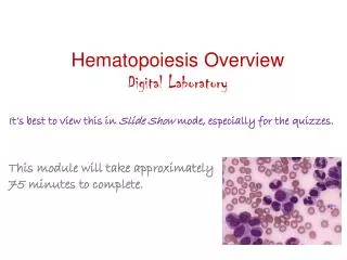
Hematopoiesis: White Blood Cells and Platelets Digital Laboratory It’s best to view this in Slide Show mode, especially for the quizzes. This module will take approximately 75 minutes to complete.
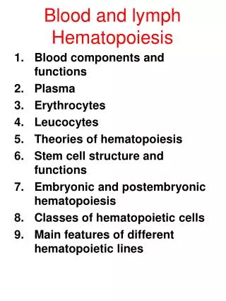
After completing this exercise, you should be able to: • Distinguish, at the light microscope level, each of the following:: • In sections of bone marrow • Red bone marrow • Yellow bone marrow • Bone marrow (or leukemia) smear • Stages of granulocyte development • Myeloblast • Promyelocyte • Myelocyte • Neutrophilicmeylocyte • Eosinophilic myelocyte • Basophilic myelocyte • Metamyelocyte • Neutrophilicmetamyelocyte • Eosinophilic metamyelocyte • Basophilic metamyelocyte • Band • Neutrophilic band cell • Eosinophilic band cell • Basophilic band cell • Mature cells (neutrophil, eosinophil, basophil) • Adipocytes • Megakaryocyte • Plasma cell • Lymphocyte
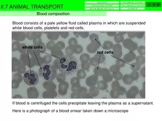
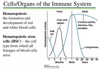
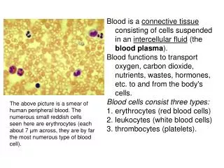
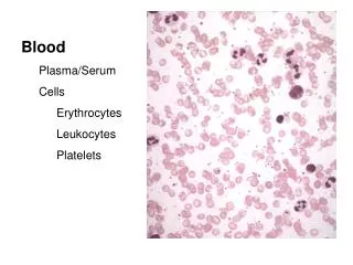
Production of blood cells – bone marrow Blood cells develop in the bone marrow from a common precursor. Once mature, these cells leave the bone marrow and enter the bloodstream, and then the tissues (note brackets at the bottom of diagram at left). We already have looked at mature blood cells in the bloodstream, as well as some of the features of developing blood cells, such as granules and nuclear shape. The next two modules focus on identification of cells that normally reside in the bone marrow.
Production of blood cells – bone marrow First, let’s focus on identifying bone marrow as a tissue. There are two types of bone marrow: --red bone marrow is shown on the left, and contains developing and mature blood cells and adipose tissue --yellow bone marrow is mostly adipose tissue If you recall the tips from the previous module, such as precursor cells are large, granules distinguish granulocytes, nuclear shape varies thru development, etc., recognizing this image as taken from red bone marrow is relatively easy. A = adipose M = megakaryocyte ( produces platelets) Ey = erythrocytes E = developing eosinophils BN = developing neutrophils
Production of blood cells – bone marrow Video of red bone marrow in situ – SL39 Video of red bone marrow in situ – SL41 • Link to SL 039 and SL 041 • Be able to identify: • Red bone marrow • Hematopoietic cells • Adipose • Yellow bone marrow (we don’t have a slide, but it’s essentially adipose in the marrow cavity)
HEMATOPOIESIS The image to the left shows the major stages of blood cell development that we will identify in these modules: The red blood cell series is shown in the first column and will be covered in the next module when we study red blood cells. The granulocyte series is the next set of columns, and will be the main focus of this module. The agranulcytes, monocytes and lymphocytes, do not have developmental stages that are histologically distinct (i.e. they are not in stages as indicated for RBCs and granulocytes on the left). Therefore, we only ask you to identify their mature forms. Distinguishing megakaryoblasts from megakaryocytes is challenging; therefore, we only ask you to identify megakaryocytes - this will be covered in this module. Again, recalling the previous module, you should recognize some of those features you studied in the cells in this image.
HEMATOPOIESIS - granulocytes In the granulocyte series, the first two recognizable stages, the myeloblast and promyelocyte, are large cells with a basophilic cytoplasm, and a large, round nucleus with a fine chromatin pattern and nucleoli . They can be distinguished from each other because the promyelocyte has non-specific (azurophilic, primary) granules, while the myeloblast does not. At these early stages, it cannot be determined in routine smears whether the myeloblast or promyelocyte is committed to the neutrophilic, eosinophilic, or basophilic cell lineage. (This can be determined with cell-type-specific markers, but that is beyond the scope of this module. Since you can’t tell whether a myeloblast or promyelocyte will become a neutrophil or eosinophil or basophil on our slides, calling a cell something like a “basophilic myeloblast” or a “neutrophilicpromyelocyte” is not advisable.
HEMATOPOIESIS - granulocytes Myeloblast --large cell --large round nucleus --nucleoli (at least one) --fine chromatin pattern --basophilic cytoplasm --absence of granules If you don’t readily recognize these features in this cell, you should return to the Hematopoiesis: Overview module.
HEMATOPOIESIS - granulocytes Note that this cell has many azurophilic granules in the cytoplasm “over” the nucleus; this obscures the fine chromatin pattern characteristic of a cell at this early stage. promyelocyte --large cell --large round nucleus --nucleoli (at least one) --fine chromatin pattern --basophilic cytoplasm --azurophilic granules --more often than not, larger than a myelocyte I’ve highlighted the major features that changed from the previous stage (i.e. how this cell differs from the myeloblast). I’ll do this for the remaining cells as well.
HEMATOPOIESIS - granulocytes Not happy with the nucleoli in these cells? Upset by the nucleus that is not so round in the cell to the right? Disappointed that the cell to the right has a little eosinophilia mixed in with the basophilia in the cytoplasm? Because development is a continuum, it is a challenge to capture “textbook” examples that exhibit every one of the useful features for the multiple views of each cell type. When I scan, I see something that looks good, and get VERY excited, only to find that the nucleolus or granules are not perfect. You can still give the cell a name, but from a teaching perspective, it’s quite challenging to find these little gems. If you doubt this, stop by my office and I’ll challenge you to a cell-finding contest; what could be more exciting than that? Fortunately for you, when we do see textbook examples, we save them for exams.
HEMATOPOIESIS - granulocytes Video of myeloblasts and promyelocytes – SL68 • Link to SL 068and SL 177 • Be able to identify: • Myeloblast • Promyelocyte
HEMATOPOIESIS - granulocytes Shown here are a neutrophilicmyelocyte (NM) and an eosinophilic myelocyte (EM). And, yes, if you are going to call a cell a myelocyte, you better say which one. NM EM myelocyte --medium-sized cell --medium-sized round nucleus --no nucleoli --clumped chromatin pattern --(mostly) basophilic cytoplasm --azurophilic granules --specific granules What actually changed from the promyelocyte are the 5 features in lighter-colored text, not the two in dark-colored text. Lots of changes at this stage.
HEMATOPOIESIS - granulocytes Video of myelocytes – SL68 • Link to SL 068and SL 177 • Be able to identify: • Myelocyte • Neutrophilicmyelocyte • Eosinophilic myelocyte • Basophilic myelocyte
HEMATOPOIESIS - granulocytes In this sequence, the nucleus changes shape: Round indented horseshoe segmented Myelocyte metamyelocyte band mature cell Nuclear shape changes are accompanied by other “minor” changes, like a general decrease in the overall size of the cell and nucleus, an even clumpier chromatin pattern, and an increase in specific granules. The most recognizable feature that differentiates these stages, however, is nuclear shape. The eosinophil and basophil series of cells also undergo similar maturation of their nuclei. These cells are not as common, however, and their large granules usually obstruct the nucleus. Therefore, we show the neutrophil series as our example here.
HEMATOPOIESIS - granulocytes Video of later granulocyte stages – SL68 • Link to SL 068and SL 177 • Be able to identify: • For neutrophils, eosinophils, and basophils • Myelocyte • Metamyelocyte • Band • Mature Note, if the presence of specific granules in eosinophils or basophils obscures the nucleus, it may be difficult to determine the exact stage of the cell.
HEMATOPOIESIS - granulocytes Before we leave granulocyte development, it should be emphasized that all cell development is a continuum, and the stages we describe here are a way for humans to organize the content, similar to stages of mitosis. Obviously, many cells will be between stages. For example, here is a cell we’ll call a promyelocyte – large cell, large round nucleus, fine chromatin with two nucleoli (white arrows), and azurophilic granules. If you look closely, however, you will see cytoplasmic eosinophilia in the Golgi region of this cell (dotted outline). Obviously, this is due to the production of neutrophilic specific granules, which is a feature of the myelocyte stage and not the promyelocyte. We call this occurrence “nuclear-cytoplasmic mismatch”. This is often striking in leukemias.
HEMATOPOIESIS - MEGAKARYOCYTES Cells that produce platelets progress through developmental stages that include progenitor cells, the megakaryoblast, and megakaryocyte. Once the megakaryocyte is formed, it resides in the marrow, next to the sinuses, where it “sheds” portions of its cytoplasm and plasma membrane into the bloodstream. These released cell fragments of the megakaryocyte are the mature platelets. A single megakaryocyte will produce thousands of platelets. The early stages (progenitor, blasts) are difficult to definitively recognize. The mature megakaryocyte, however, is easily recognized in bone marrow.
HEMATOPOIESIS - MEGAKARYOCYTES During maturation, megakaryoblasts duplicate their genome (i.e. 2N 4N 8N 16N 32N 64N ) without nuclear division. In addition, the cell expands its basophilic cytoplasm, which will be “shed” to produce platelets. The resulting megakaryocytes are big cells, with extensive eosinophilic cytoplasm, often foamy, with granules and a multi-lobed nucleus. This image from a bone marrow smear shows a single, REALLY bigmegakaryocyte (outlined, compare to neighboring RBCs), with nice lobulation of the nucleus. The cytoplasm shows some remaining basophilia, but you can clearly see eosinophilic regions as well.
HEMATOPOIESIS - MEGAKARYOCYTES This image was taken from a red bone marrow section (not a smear, but a section of a bone), and is magnified only to 40X. The numerous nuclei observed throughout the slide represent different stages of blood cell development – but staging is not routinely accomplished in such material because the magnification is insufficient, and because the features of each cell are obscured by both the superimposition of cells in the thickness of a routine section and the proximity of cells to one another. Despite this, two megakaryocytes are easily identified - large cells, with multi-lobed nuclei and eosinophilic cytoplasm.
HEMATOPOIESIS - MEGAKARYOCYTES Video of megakaryocyte in a bone marrow smear – SL68B Video of megakaryocyte in a bone marrow tissue – SL41 • Link to SL 068B and SL 041 • Be able to identify: • megakaryocyte
Lymphocytes and plasma cells You saw these cells in the quiz in the hematopoiesis overview module. Don’t forget that you will see lymphocytes and plasma cells in bone marrow. You will see mature granulocytes as well.
After completing this exercise, you should be able to: • Distinguish, at the light microscope level, each of the following:: • In sections of bone marrow • Red bone marrow • Yellow bone marrow • Bone marrow (or leukemia) smear • Stages of granulocyte development • Myeloblast • Promyelocyte • Myelocyte • Neutrophilicmeylocyte • Eosinophilic myelocyte • Basophilic myelocyte • Metamyelocyte • Neutrophilicmetamyelocyte • Eosinophilic metamyelocyte • Basophilic metamyelocyte • Band • Neutrophilic band cell • Eosinophilic band cell • Basophilic band cell • Mature cells (neutrophil, eosinophil, basophil) • Adipocytes • Megakaryocyte • Plasma cell • Lymphocyte
Final quiz Self-check: Identify the cell indicated by the arrow. (advance slide for answer) neutrophilic band (stab) cell
Final quiz Self-check: Identify the cell indicated by the arrow. (advance slide for answer) promyelocyte
Final quiz Self-check: Identify the cell indicated by the arrow. (advance slide for answer) Eosinophilic band cell
Final quiz Self-check: Identify the cell indicated by the arrow. (advance slide for answer) promyelocyte
Final quiz Self-check: Identify the cells indicated by the arrow. (advance slide for answer) Neutrophilicmetamyelocyte
Final quiz Self-check: Identify the cell indicated by the arrow. (advance slide for answer) Basophil or a precursor basophil (e.g. basophilic band cell or basophilic metamyelocyte)
Final quiz Self-check: Identify the cell indicated by the arrow. (advance slide for answer) lymphocyte
Final quiz Self-check: Identify the cell indicated by the arrow. (advance slide for answer) Eosinophilic metamyelocyte
Final quiz Self-check: Identify the cell indicated by the arrow. (advance slide for answer) Plasma cell
Final quiz Self-check: Identify the cell indicated by the arrow. (advance slide for answer) Eosinophilic myelocyte
Final quiz Self-check: Identify the cell indicated by the arrow. (advance slide for answer) megakaryocyte
Final quiz Self-check: Identify the cell indicated by the arrow. (advance slide for answer) myeloblast
Final quiz Self-check: Identify the cell indicated by the arrow. (advance slide for answer) Neutrophilicmyelocyte
Final quiz Self-check: Identify the cell indicated by the arrow. (advance slide for answer) Neutrophilicmyelocyte The cytoplasm in this neutrophil form looks highly eosinophilic but gain clues to the true ID by looking around at the other obvious neutrophil forms and the degree of eosinophilia characteristic of them on this particular smear.
Final quiz Self-check: Identify the cell indicated by the arrow. (advance slide for answer) Neutrophilic band (stab) cell
Final quiz Self-check: Identify the cell indicated by the arrow. (advance slide for answer) Eosinophilic metamyelocyte
Final quiz Self-check: Identify the cell indicated by the arrow. (advance slide for answer) Basophil or a precursor basophil (e.g. basophilic band cell or basophilic metamyelocyte)
Final quiz Self-check: Identify the cell indicated by the arrow. (advance slide for answer) eosinophil
Final quiz Self-check: Identify the cell indicated by the arrow. (advance slide for answer) (Nearly mature) neutrophil
Final quiz Self-check: Identify the cell indicated by the arrow. (advance slide for answer) Eosinophilic myelocyte
Final quiz Self-check: Identify the cell indicated by the arrow. (advance slide for answer) eosinophil
Final quiz Self-check: Identify the cell indicated by the arrow. (advance slide for answer) myeloblast
Final quiz Self-check: Identify the cell indicated by the arrow. (advance slide for answer) Plasma cell
Final quiz Self-check: Identify the cell indicated by the arrow. (advance slide for answer) promyelocyte