Download
1 / 3
50 likes | 202 Vues
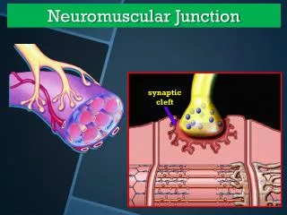
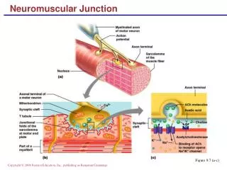
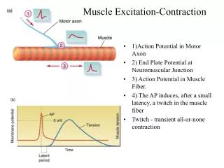
The signal for contraction comes down the axon of a motor neuron to the neuromuscular junction. When it arrives at the axon terminal, the synaptic vesicles fuse with the membrane and release the neurotransmitter (acetylcholine) into the synaptic cleft.

E N D
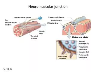
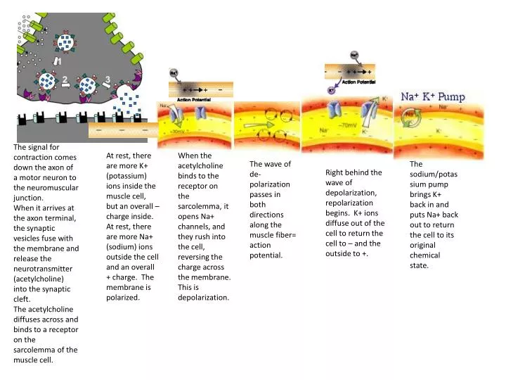
The signal for contraction comes down the axon of a motor neuron to the neuromuscular junction. When it arrives at the axon terminal, the synaptic vesicles fuse with the membrane and release the neurotransmitter (acetylcholine) into the synaptic cleft. The acetylcholine diffuses across and binds to a receptor on the sarcolemma of the muscle cell. At rest, there are more K+ (potassium) ions inside the muscle cell, but an overall – charge inside. At rest, there are more Na+ (sodium) ions outside the cell and an overall + charge. The membrane is polarized. When the acetylcholine binds to the receptor on the sarcolemma, it opens Na+ channels, and they rush into the cell, reversing the charge across the membrane. This is depolarization. The wave of de-polarization passes in both directions along the muscle fiber= action potential. The sodium/potassium pump brings K+ back in and puts Na+ back out to return the cell to its original chemical state. Right behind the wave of depolarization, repolarization begins. K+ ions diffuse out of the cell to return the cell to – and the outside to +.
The action potential passes into the muscle fiber through the T (transverse) tubules. The cisterna of the SR (sarcoplasmic reticulum) are stimulated to release Ca++ (calcium ions). These ions diffuse to the sarcomeres where the Ca++ attaches to the troponin on the thin myofilament.
The binding of the Ca++ to the troponin causes the tropomyosin to pull out of the way, exposing binding sites for the attachment of the myosin heads to the actin. The myosin heads bend and pull on the actin filaments, bringing them closer together and shortening the sarcomere. The heads release and recock, then reattach and bend and pull. This process continues until the sarcomere reaches full contraction. When acetylcholinesterase breaks down acetylcholine, the stimulus to the muscle stops. Ca++ is actively reabsorbed by the sarcoplasmic reticulum. Without Ca++, the tropomyosin blocks the binding sites so that the myosin heads cannot attach. The filaments release and relax. Contraction ceases.