
The Lower Torso
E N D
Presentation Transcript
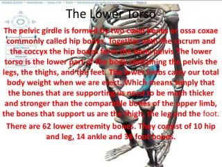
The Lower Torso The pelvic girdle is formed by two coxal bones or ossacoxae commonly called hip bones. Together with the sacrum and the coccyx the hip bones form the bony pelvis The lower torso is the lower part of the body containing the pelvis the legs, the thighs, and the feet. The lower limbs carry our total body weight when we are erect. Which means simply that the bones that are supporting us need to be much thicker and stronger than the comparable bones of the upper limb, the bones that support us are the thigh, the leg and the foot. There are 62 lower extremity bones. They consist of 10 hip and leg, 14 ankle and 38 foot bones.
Thigh • Femur- is the only bone in the thigh and it is also the heaviest, and the strongest bone in the body. Its proximal end has a ball-like head a neck and greater and lesser trochanters separated by the intertrochanteric line and posteriorly by the intertrochanteric crest. • The trochanters intertrochanteric crest all located on the shaft all serve as sites for muscle attachment. • Anteriorly on the distal femur is the smooth patellar surface, which forms a joint with the patella, or kneecap.
Leg The leg is technically only the region from the knee to the ankle. It is formed by the fibula on side away from the body ( lateral side) and the tibia, also called the shin bone, on the side nearest the body ( medial side). The tibia connects to the femur to form the knee joint and with the talus, a foot bone, to allow the ankle to flex and extend. The tibia is larger than the fibula because it bears most of the weight, while the fibula serves as an area for muscle attachment • The legs connected along their length by an interosseous membrane, two bones, the tibia and fibula, form the skeleton of the leg. Also at the end of the tibia the medial and lateral condyles are separated by the intercondylar eminence articulate with the distal end of the femur to form the knee joint. • The fibula lies alongside the tibia but has absolutely nothing to do with forming the knee joint but it forms the outer part of the ankle.
Foot • The foot, or pes, contains the 26 bones of the ankle, instep, and the five toes. The ankle, or tarsus, is composed of the 7 tarsal bones which correspond to the carpals in the wrist. The largest tarsal bone is called the calcaneus or heel bone. The talus rests on top of the calcaneus and is connected to the tibia. Directly in front of the talus is the navicular bone. The remaining bones from medial to lateral are the medial, intermediate, the lateral cuneiform bones, and the cuboid bone. The metatarsal and phalanges bones of the foot are similar in number and position to the metacarpal and phalanges bones of the hand. The five metatarsal bones are numbered I to V starting on the medial side with the big toe. The first metatarsal bone is larger than the others because it plays a major role in supporting the body's weight. The 14 phalanges of the foot, as with the hand, are arranged in a proximal row, a middle row, and a distal row, with the big toe, or hallux, having only a proximal and distal phalanx. The foot's two arches are formed by the structure and arrangement of the bones and are maintained by tendons and ligaments. The arches give when weight is placed on the foot and spring back when the weight is lifted off of the foot. The arches may fall due to a weakening of the ligaments and tendons in the foot.
Foot Cont. • The foot which is composed of the tarsal's, the metatarsals, and phalanges have two important functions. 1 it supports our body weight , 2 it serves as a lever that allows us to propel our bodies forward when we walk and run.
The Patella(Knee-Cap) • The patella or kneecap is a large, triangular sesamoid bone between the femur and the tibia. It is formed in response to the strain in the tendon that forms the knee. The patella protects the knee joint and strengthens the tendon that forms the knee. The bones of the lower extremities are the heaviest, largest, and strongest bones in the body because they must bear the entire weight of the body when a person is standing in the upright position.
Male and Female Pelvis Comparisons • The female pelvis as a whole is shallower, and the bones are lighter and thinner. • The female ilia flare more laterally. • The female sacrum is shorter and less curved • The female ischial spines are shorter and farther apart: thus the outlet is larger • The female pubic arch is more rounded because the angle of the pubic arch is greater.
References • Anatomy Textbook