Download
1 / 78
790 likes | 1.14k Vues
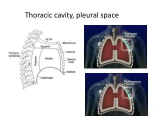
Physiology of the Pleural Space . Visceral Pleura. Covers the lung parenchyma including the interlobar fissures. Provides mechanical support to the lung. Limits lung expansion, protecting the lung . Contributes to the elastic recoil of the lung and lung deflation. . Visceral Pleura.

E N D
Visceral Pleura • Covers the lung parenchyma including the interlobar fissures. • Provides mechanical support to the lung. • Limits lung expansion, protecting the lung . • Contributes to the elastic recoil of the lung and lung deflation.
Visceral Pleura • Thick visceral pleura: mesothelium and dense layer of connective tissue. • Systemic circulation. Bronchial arteries. • Blood, lymph vessels and nerves. • Humans, sheep, cows, pigs and horses. • Thin visceral pleura: monkeys, dogs and cats. • Pulmonary circulation.
Pleural Visceral • Mean thickness:25-83 um. • Distance from the microvessels to the pleura: 18-56um. • Drainage into the pulmonary veins.
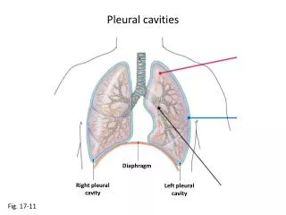
Parietal Pleura • Lines the inside of the thoracic cavities. • Subdivided into the costal, mediastinal and diaphragmatic parietal pleura. • Loose connective tissue and single layer of mesothelial cells. • Capillaries and lymphatic lacunas. • Blood supply from systemic capillaries. • Blood drainage: Intercostal veins.
Parietal Pleura • Mean thickness : 20-25 um. • Distance from the micro vessel to the pleural space: 10-12 um. • Stomas communicates lymphatic vessels with pleural space. • Nitric oxide. • Diaphragmatic pleura.
Parietal Pleura • Lymphatic lacunas are in communication with the pleural space by stomas and ultimately drain to the internal mammary, para aortic and diaphragmatic lymph nodes.
Mesothelial Cells • Very active cells. • Mesothelium. Dynamic cellular membrane. • Transport and movement of fluid and particulate matter. • Leukocyte migration. • Synthesis of cytokines, growth factors and extracellular proteins.
Mesothelial Cells • Mesothelial cell can convert to macrophages and myofibroblasts. • TGF Beta. • Microvilli: entangle glycoproteins rich in hyaluronic acid.
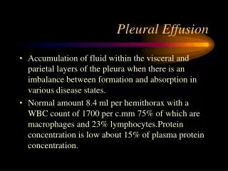
Pleural Fluid • 8.4 +/- 4.3 mL. • Total pleural fluid volume: 0.26 +/- 0.1 mL/Kg. • Cell count: WBC: 1716x103cells/ml. RBC: 700x103 cells/ml. • Macrophages:75%. • Lymphocytes: 23% • Mesothelial cells, neutrophils and eosinophils:2%.
Pleural Pressure • Negative pressure generated between the visceral and parietal pleura by the opposing elastic forces of the chest wall and lung at FRC. • Represents the balance between the outward pull of the thoracic cavity and the inward pull of the lung.
Pleural Pressure • It is the pressure at the outer surface of the lung and the heart and inner surface of the thoracic cavity. • Distensible structures. • Compliance and pressure difference between inside and outside. • Primary determinant of the lung, cardiac and thoracic cavity volume.
Measurement • Indirect measurement. Esophageal pressure. • Lower one third of the esophagus. • Upright posture. • Analysis of lung and chest wall compliance, work of breathing, respiratory muscle function and the presence of diaphragm paralysis. • Mechanical ventilation guided by esophageal pressures in ALI. November 13, 2008.
Pleural Pressure • Pleural pressure is not uniform. • Gradient between the superior ( lowest, most negative) and inferior (highest, least negative) portions of the lung. • 0.3 cm H20/ cm vertical distance. • In the upright position, gradient of pleural pressure between the apex and the base is approximately 8 cm H20.
Pleural Pressure • Gravity. • Mismatching of the shapes of the lung and chest wall. • The weight of the lung and other intra thoracic structures.
Alveolar pressure is constant throughout the lung. • Differents parts of the lung have different distending pressures.
Pleural Pressure • Different parts of the lungs have different distending pressures. • The alveoli in the superior parts of the lung tends to be larger than those in the inferior parts. • Formation of pleural blebs. • Uneven distribution of ventilation.
Pleural fluid formation • Pleural capillaries. • Interstitial space in the lung. • Intra thoracic blood vessels. Hemothorax. • Intra thoracic lymphatics. Chylothorax. • Peritoneal cavity.
Starling’s Law of Trans capillary Exchange Qf = Lp x A[(Pcap-Ppl) –σd(πcap-πpl)] Qf= liquid movement Lp = filtration coefficient /unit area of the membrane A = surface area of the membrane σd = solute reflection coefficient for protein (membrane's ability to restrict passage of large molecules P = hydrostatic pressures π = oncotic pressure
Pleural Capillaries • A gradient for fluid formation is normally present in the parietal pleura. • Hydrostatic pressure: 30cm H20. • Pleural Pressure: -5 cm H20. • Oncotic pressure in plasma: 34 cm H20. • Oncotic pressure in the pleural fluid: 5 cm H20. • Net pressure gradient: 6 cm H20.
Pleural Capillaries • Net gradient: close to zero. • Pleural visceral capillaries drain into the pulmonary veins. • The filtration coefficient is substantially less than for the parietal pleura.
Interstitial origin • Much of the pleural fluid. • High pressure pulmonary edema: the pleural fluid formed is directly related to the elevation in the wedge pressure. • Increases in pleural fluid formation occurs only after the development of pulmonary edema.
The presence of pulmonary effusion is more closely correlated with the pulmonary venous pressure than with the systemic venous pressure. • High permeability pulmonary edema: Pleural fluid accumulates only after pulmonary edema develops.
In general : pleural effusion develops when the extravascular lung water has reached a critical level in a certain amount of time. • 5-8 g of fluid/ gram of dry lung.
Increasing levels of interstitial fluid is related with increase in sub pleural interstitial pressure, allowing fluid transverse the visceral pleura to the pleural space. • Pressure gradient rise from 1.3 to 4.4 cm H2O. • Associated to rise in lung water to 5-6 g/g dry lung.
Peritoneal Cavity • Free fluid in the peritoneal cavity. • Opening in the diaphragm. Diaphragmatic defects. • Pressure in the pleural cavity is less than the pressure in the peritoneal cavity.
Pleural Fluid Absorption • Mean lymphatic flow is 0.22-0.4 mL/kg/hour. • Lymphatics operate at maximum capacity once the volume of the pleural liquid exceeds a certain threshold. • The capacity for lymphatic clearance is 28 times as high as the normal rate of pleural fluid formation.
Pathogenesis of Pleural Effusion • Pleural fluid formation exceeds the rate of pleural fluid absorption.
Effects of Pneumothorax on PleuralPressure • Air will flow into the pleural space until a pressure gradient no longer exits or until the communication is sealed.
Effects of Pneumothorax on Pleural Pressure • The distribution in the increase in the pleural pressure is homogenous and the pressure is the same throughout the entire pleural space. • In pleural effusion there is a gradient in the pleural pressure due to the hydrostatic column of fluid.
Effects of Pneumothorax on Pleural Pressure • The upper lobe is affected more than the lower lobe in pneumothorax, because the pressure in the apices is much more negative that at the bases.
Effects of Pneumothorax in Lung Function • Restrictive ventilatory defect. • Decreased Vital Capacity. • Decreased FRC. • Decreased TLC. • Decreased RV. • Decreased end expiratory lung volume. • Increased end expiratory thoracic volume?. • Slightly decreased DLCO.