Download
1 / 1
20 likes | 190 Vues
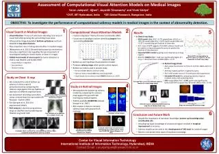
Assessment of Computational Visual Attention Models on Medical Images. Varun Jampani 1 , Ujjwal 1 , Jayanthi Sivaswamy 1 and Vivek Vaidya 2 1 CVIT, IIIT Hyderabad, India 2 GE Global Research, Bangalore, India.

E N D
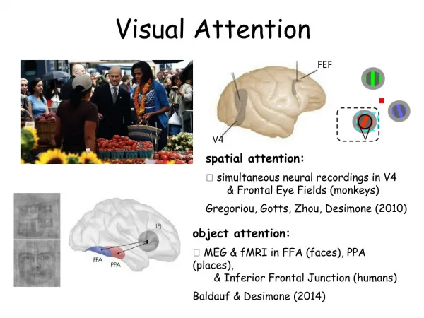
Assessment of Computational Visual Attention Models on Medical Images Varun Jampani1, Ujjwal1, Jayanthi Sivaswamy1 and Vivek Vaidya2 1CVIT, IIIT Hyderabad, India 2GE Global Research, Bangalore, India • OBJECTIVE: To investigate the performance of computational saliency models in medical images in the context of abnormality detection. 1 2 5 • Results • Computational Visual Attention Models Visual Search in Medical Images • Feature Integration Theory [Treisman and Gelade 1980] • Visual search paradigm involves identifying targetsfrom surrounding distractors • Visual Attention: Process of selectively attending to an area of visual field while ignoring the surrounding visual areas • Influenced by image-dependent bottom-up features and task-dependent top-down features • Plays important role in finding abnormalities in medical images • [Matsumoto et al. 2011]: Showed that bottom-up mechanisms also play a significant role in guiding the eye movements of neurologists looking for stroke lesions on brain CT images • Global-Focal model of visual search for tumor detection in chest x-rays [Nodine and Kundel 1987] • Overall Pattern recognition • Focal attention • Decision making • In Chest X-ray Study • GBVS model (Mdn AUC = 0.77) outperforms (Z=0.0, p < .001) SR model (Mdn AUC = 0.67) and IK model (Mdn AUC = 0.67 ) in predicting eye fixations of observers • AUC value of 0.77 suggests that GBVS saliency model can be used to a reasonably good accuracy to predict the fixations of the observers • Extended GBVS model by giving more importance to lung regions • AUCs for EGBVS (Mdn = 0.81) are significantly higher (Z = 2.0, p < .001) than those of GBVS (Mdn = 0.77) Mean AUCs corresponding to different saliency models for all the observers in chest x-ray study Disjunctive Target Conjunctive Target • Bottom-up and Top-Down Visual attention models • Produces saliency maps which predict salient regions • Bottom-up models used in present study • Itti-Koch Model (IK) [Itti and Koch 2001] • Spectral residual model (SR) [Hou and Zhang 2007] • Graph based visual saliency model (GBVS) [Harel et al. 2007] • In Retinal Image Study • AUC values found to be: 0.72 for IK, 0.70 for GBVS and 0.73 for SR • All three models perform roughly the same • AUC of SR model rose to 0.74 with optic disk suppression • Extended SR model by computing saliency maps at multiple scales and combining them • ESR model (Mean AUC = 0.94) performed significantly better (28.76% increase) than SR model (Mean AUC = 0.74) 3 • Study on Chest X-rays • We evaluated the role of bottom-up saliency in chest x-rays of pneumoconiosis by comparing the saliency maps against the eye fixations of observers of different expertize levels. • Eye movement recordings were done using a remote head free eye tracker (SR Research - Eyelink 1000) • See [Jampani et al. 2011] for experimental details • ROC analysis is done by using saliency maps as classifiers and considering eye fixations as ground truth 4 Average ROC curves for different saliency models in retinal image study • Study on Retinal Images • We evaluated the bottom-up saliency models by comparing the saliency maps against the ground truth markings by medical experts • Publicly available DIARETDB1 dataset [Kauppi et al. 2007] • ROC analysis is done to compare saliency maps with ground truth markings Input Image Sample X-ray segments showing different stages of Pneumoconiosis (Conjunctive Targets) Itti-Koch GBVS Different steps in attracting EGBVS saliency map from a sample chest x-ray Steps for deriving ESR saliency maps SR 6 Saliency maps extracted from 2 sample retinal images • Conclusion and Future Work Eye fixations of an observer on a sample chest x-ray • Despite the importance of top-down knowledge, bottom-up knowledge plays considerable role • Including top-down knowledge of anatomical regions resulted in marginal improvement in accuracy • Saliency models can be used in the development of CAD tools for medical images • Need to incorporate other types of top-down knowledge A sample retinal image and ground truth markings by four medical experts. Taken from DIARETDB1 dataset [Kauppi et al. 2007] Sample retinal images showing hard exudates (Disjunctive Targets) A sample chest x-ray and the corresponding saliency maps computed using different saliency models Center for Visual Information Technology International Institute of Information Technology, Hyderabad, INDIA Contact Email: varunjampani@research.iiit.ac.in