Complicated Case of Lupus Nephritis with Thromboembolic Events and Lung Lesions
160 likes | 289 Vues
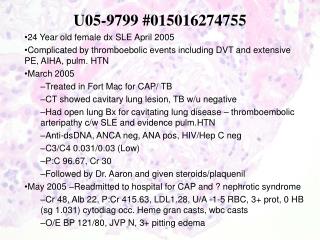
This case study highlights a 24-year-old female diagnosed with systemic lupus erythematosus (SLE) in April 2005, experiencing multiple complications including thromboembolic events (DVT and extensive pulmonary embolism), autoimmune hemolytic anemia, and pulmonary hypertension. Initial treatment included hospitalization for community-acquired pneumonia and an extensive workup for cavitary lung lesions, which tested negative for tuberculosis. A renal biopsy diagnosed mild activity mesangial proliferative lupus nephritis, revealing significant mesangial staining patterns.

Complicated Case of Lupus Nephritis with Thromboembolic Events and Lung Lesions
E N D
Presentation Transcript
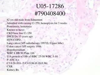
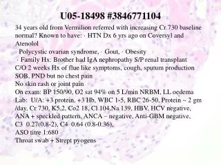
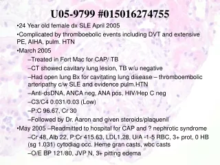
U05-9799 #015016274755 • 24 Year old female dx SLE April 2005 • Complicated by thromboebolic events including DVT and extensive PE, AIHA, pulm. HTN • March 2005 • Treated in Fort Mac for CAP/ TB • CT showed cavitary lung lesion, TB w/u negative • Had open lung Bx for cavitating lung disease – thromboembolic arteripathy c/w SLE and evidence pulm.HTN • Anti-dsDNA, ANCA neg, ANA pos, HIV/Hep C neg • C3/C4 0.031/0.03 (Low) • P:C 96.67, Cr 30 • Followed by Dr. Aaron and given steroids/plaquenil • May 2005 –Readmitted to hospital for CAP and ? nephrotic syndrome • Cr 48, Alb 22, P:Cr 415.63, LDL1.28, U/A -1-5 RBC, 3+ prot, 0 HB (sg 1.031) cytodiag occ. Heme gran casts, wbc casts • O/E BP 121/80, JVP N, 3+ pitting edema
IF • IgG- Moderate mesangial staining • IgA- Mild-moderate mesangial staining • IgM- Moderate mesangial staining • C3- Mild mesangial staining • C1q- Moderate mesangial and vascular staining • Kappa- Mild mesangial staining • Lambda- Moderate mesangial staining • Fibrin- Moderate interstitial staining • Albumin- Mild hyalen droplet change in tubular cytoplasm
Diagnosis:Renal Biopsy:Mesangial proliferative lupus nephritis of mild activity and no chronicity